Comparative Study
. 2005 Jul;12(7):615-8.
doi: 10.1038/nsmb955. Epub 2005 Jun 19.

Cryo-electron microscopy reconstruction of a poliovirus-receptor-membrane complex

Affiliations
Comparative Study

Cryo-electron microscopy reconstruction of a poliovirus-receptor-membrane complex

Doryen Bubeck et al. Nat Struct Mol Biol. 2005 Jul.

Abstract

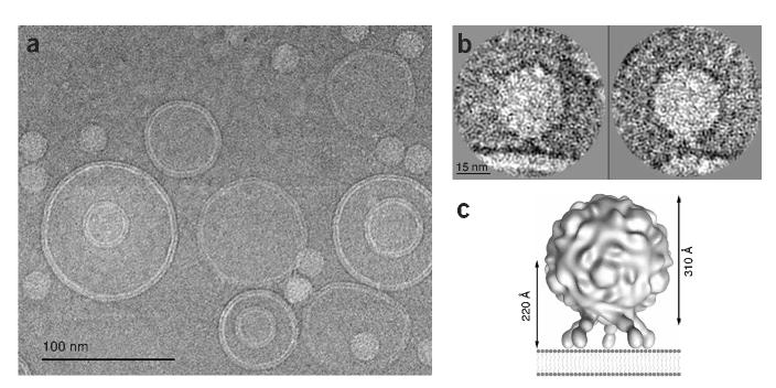
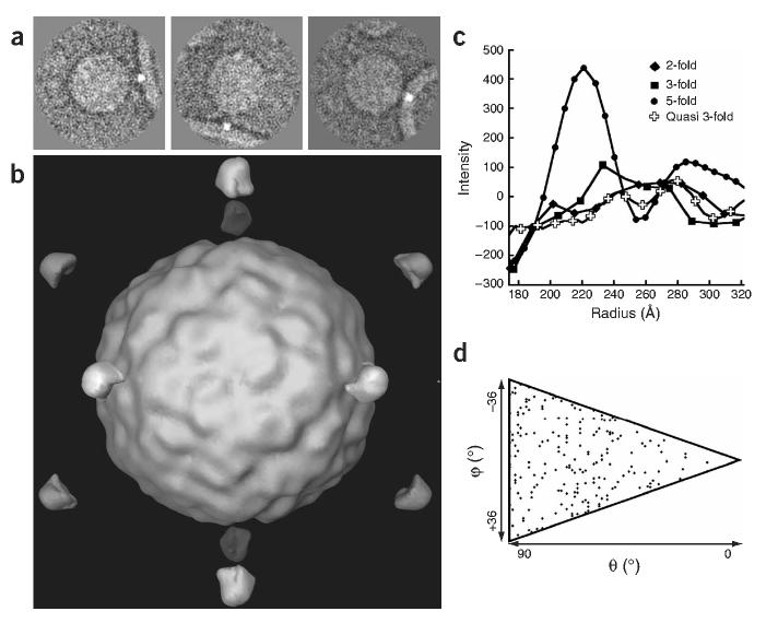
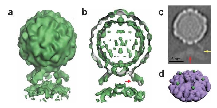
To study non-enveloped virus cell entry, a versatile in vitro model system was developed in which liposomes containing nickel-chelating lipids were decorated with His-tagged poliovirus receptors and bound to virus. This system provides an exciting opportunity for structural characterization of the early steps in cell entry in the context of a membrane. Here we report the three-dimensional structure of a poliovirus-receptor-membrane complex solved by cryo-electron microscopy (cryo-EM) at a resolution of 32 A. Methods were developed to establish the symmetry of the complex objectively. This reconstruction demonstrates that receptor binding brings a viral five-fold axis close to the membrane. Density is clearly defined for the icosahedral virus, for receptors (including known glycosylation sites) and for the membrane bilayer. Apparent perturbations of the bilayer close to the viral five-fold axis may function in subsequent steps of cell entry.


Figures

Figure 1
Figure 1
Poliovirus bound to receptor-decorated liposomes as visualized by cryo-EM. (a) Electron micrograph field showing a fairly uniform population of receptor-decorated liposomes and multiple instances of virus attachment. (b) Individually windowed particles in which density corresponding to receptor can be observed linking native virus to the membrane. (c) Model-based electron density was calculated from pseudo atomic models fitted by Belnap et al. into a cryo-EM reconstruction of receptor-decorated poliovirus. In this diagram, the distance between the virus center and the nearest membrane contact point (assuming a planar bilayer) is predicted to be 220Å for an approach along the viral five-fold axis.
Figure 2
Figure 2
Artificially dotted icosahedral reconstruction of a virus-receptor-liposome complex demonstrates that virus approaches membrane along its five-fold axis. (a) Two-dimensional images in various orientations were modified by adding dots to the liposome position closest to the virus. (b) Three-dimensional reconstruction computed from dotted images using icosahedral orientations from undotted images. (c) Density values sampled along various radial directions, measured from the virus center. The peak value at 221Å along the five-fold axis is significantly higher than in other directions. (d) Distribution of particle orientations in the final five-fold symmetric reconstruction. Each diamond represents one usable two-dimensional image in the space of unique orientations (triangle).
Figure 3
Figure 3
Five-fold symmetric three-dimensional reconstruction of native poliovirus bound to receptor-decorated liposomes. (a) In a surface rendering of the complex (green), the icosahedral features of the virus capsid are maintained, and the density for receptors and membrane are well defined. (b) A cut-away view shows that the three-dimensional reconstruction of the complex (green) superimposes well on electron density (gray) corresponding to the virus-receptor complex in solution. Extra density on the outer leaflet of the membrane near the viral five-fold axis could be due to a perturbation of the membrane. A red arrow indicates a glycosylation site on domain 2 of Pvr. (c) A gray-scale representation of a central slice of the reconstruction that is 27Å thick. This view shows the protrusion of the membrane toward the viral five-fold axis (red arrow) and a weak but continuous density connection between the C-terminal domain of the receptor and the membrane (yellow arrow). Note the clear separation between the inner and outer leaflets of the bilayer, in both the bulged and peripheral areas of the membrane. (d) An unobstructed view of the membrane (purple) shows the crownlike appearance of the apparent distortion. Before removing the receptor density, the small green spheres were located near the predicted C terminus of receptor domain 3. For clarity, d shows lower contour levels than a and b.

References

    1. Hogle JM, Chow M, Filman DJ. Three-dimensional structure of poliovirus at 2.9Å resolution. Science. 1985;229:1358–1365. - PubMed
    1. Mendelsohn CL, Wimmer E, Racaniello VR. Cellular receptor for poliovirus: molecular cloning, nucleotide sequence, and expression of a new member of the immunoglobulin superfamily. Cell. 1989;56:855–865. - PubMed
    1. Selinka HC, Zibert A, Wimmer E. Poliovirus can enter and infect mammalian cells by way of an intercellular adhesion molecule 1 pathway. Proc. Natl. Acad. Sci. USA. 1991;88:3598–3602. - PMC - PubMed
    1. Tosteson M, Wang H, Naumov A, Chow M. Poliovirus binding to its receptor in lipid bilayers results in particle-specific, temperature-sensitive channels. J. Gen. Virol. 2004;86:1581–1589. - PubMed
    1. Lonberg-Holm K, Gosser LB, Kauer JC. Early alteration of poliovirus in infected cells and its specific inhibition. J. Gen. Virol. 1975;27:329–345. - PubMed

Publication types

MeSH terms