. 2005 Jul;115(7):1797-805.
doi: 10.1172/JCI24176. Epub 2005 Jun 16.

Chronic lymphocytic leukemia cells induce changes in gene expression of CD4 and CD8 T cells

Affiliations

Chronic lymphocytic leukemia cells induce changes in gene expression of CD4 and CD8 T cells

Güllü Görgün et al. J Clin Invest. 2005 Jul.

Abstract

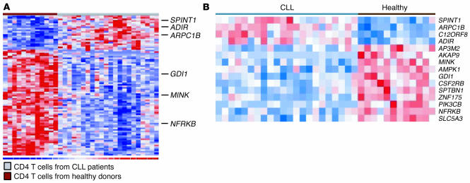
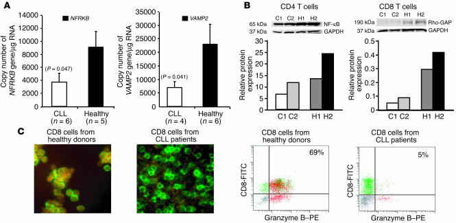
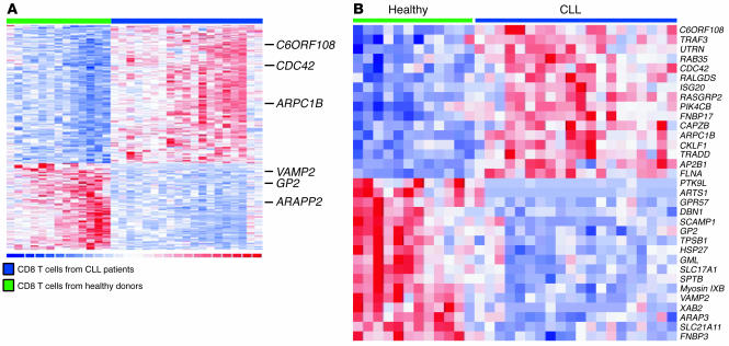
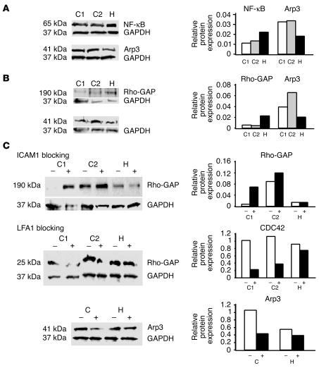
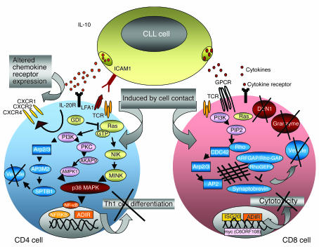
To examine the impact of tumors on the immune system, we compared global gene expression profiles of peripheral blood T cells from previously untreated patients with B cell chronic lymphocytic leukemia (CLL) with those from age-matched healthy donors. Although the cells analyzed were not part of the malignant clone, analysis revealed differentially expressed genes, mainly involved in cell differentiation in CD4 cells and defects in cytoskeleton formation, vesicle trafficking, and cytotoxicity in CD8 cells of the CLL patients. In coculture experiments using CLL cells and T cells from healthy allogeneic donors, similar defects developed in both CD4 and CD8 cells. These changes were induced only with direct contact and were not cytokine mediated. Identification of the specific pathways perturbed in the T cells of cancer-bearing patients will allow us to assess steps to repair these defects, which will likely be required to enhance antitumor immunity.


Figures

Figure 1
Figure 1
Differentially expressed genes in CD4 cells from patients with CLL compared with healthy donors. Dendrogram of differentially expressed genes by supervised analysis (P < 0.05). (A) CD4 cells from patients with CLL compared with healthy donors. Twenty-two genes were significantly increased (red) and 68 genes significantly decreased (blue) in CD4 cells from CLL patients. (B) Genes involved in Ras-dependent JNK and p38 MAPK pathways in CD4 cells. The dendrogram represents selected genes from A.
Figure 2
Figure 2
Validation of gene expression observed by microarray. (A) Concordant with data seen on microarray, by quantitative PCR there was decreased expression of NFRKB in CD4 cells and VAMP2 in CD8 cells from CLL patients compared with healthy donors. The figure represents data from CD4 cells from 6 CLL patients and 5 healthy donors and CD8 cells from 4 CLL patients and 6 healthy donors. Statistical significance was assessed in a 2-tailed Student’s t test. (B) Decreased expression of NF-κBp65 in CD4 cells and Rho-GAP p190 proteins in CD8 cells from CLL patients compared with healthy donors. The left 2 lanes represent protein expression in CD4 or CD8 cells from 2 CLL patients (C1 and C2), and the right 2 lanes represent 2 healthy donors (H1 and H2). The expression of proteins was normalized by GAPDH expression level and is shown as protein bands and densitometric intensity of each band. The figure is representative of 3 additional experiments performed on 6 different donors, all showing a similar pattern (P < 0.05). (C) Intracytoplasmic expression of the GP1 gene product granzyme B, detected in CD8 cells from CLL patients and healthy donors by flow cytometry and fluorescent microscopy. To obtain at least 99% CD8 cell population, cells were purified using magnetically labeled negative cell-depletion antibodies. High expression of granzyme B in CD8 cells from healthy donors (CD8-FITC+ granzyme B–PE+, orange-brown) was observed compared with that in CD8 cells from CLL patients (CD8-FITC+ granzyme B–PE, green). The figure is representative of experiments performed with 4 different patients and healthy donors (P < 0.05).
Figure 3
Figure 3
Differentially expressed genes in CD8 cells from patients with CLL. Dendrogram of differentially expressed genes by supervised analysis in CD8 cells from patients with CLL compared with healthy donors (P < 0.05). (A) One hundred sixty-eight genes were significantly increased (red) and 105 genes were significantly decreased (blue) in CD8 cells from CLL patients. (B) Dendrogram of differentially expressed genes involved in cytoskeleton formation, vesicle trafficking, and cytotoxicity pathways in CD8 cells. The dendrogram represents selected genes from A.
Figure 4
Figure 4
Impact of T cell–cancer cell contact on healthy T cells. Highly purified T cells from healthy donors were cocultured with B cells from CLL patients or from their HLA-matched healthy donors at a 2:1 (T/B cell) ratio for 48 hours. (A) Decrease in 65-kDa NF-κB and increase in 41-kDa Arp3 in healthy CD4 cells after contact with allogeneic CLL cells (C1 and C2) and healthy B cells (H). (B) Decrease in 190-kDa Rho-GAP and increase in 41-kDa Arp3 in healthy CD8 cells after contact with CLL cells (C1 and C2) and healthy allogeneic B cells (H). (C) Impact of CLL cell contact on cytoskeletal protein expression in allogeneic healthy T cells, confirmed by ICAM1 or LFA1 blocking. Highly purified CD4 or CD8 cells from healthy donors were cocultured with B cells from CLL patients (C1 and C2) or healthy donors (H) with or without blockade of the LFA1 or ICAM1 interaction. Expression of 190-kDa Rho-GAP in healthy CD8 cells was increased after blockade of the ICAM1 during CLL cell–T cell contact, and expression of 41-kDa Arp3 in CD4 T cells and 25-kDa CDC42 in healthy CD8 cells was decreased after blockade of the LFA1 or ICAM1. –, protein expression in nonblocked cells; +, protein expression in blocked cells. Protein expressions were normalized by GAPDH expression level and are shown as protein bands and densitometric intensity of each band. The figure is representative of 3 different experiments performed with 6 different patients with CLL and 6 different healthy donors showing a similar pattern (P < 0.05).
Figure 5
Figure 5
Differentially expressed genes by their involvement in specific signaling pathways. Representative defects in T cell pathways and functions caused by CLL cell–T cell contact are shown in this diagram. Differentially expressed genes involved in cell differentiation, particularly JNK (pink) and p38 MAPK (yellow) pathways and cytoskeleton formation and vesicle transportation (blue), in CD4 T cells from CLL patients compared with healthy donors are represented by selected genes that were increased (rectangles) or decreased (ovals). Differentially expressed genes involved in cytoskeleton formation, vesicle trafficking (blue), and cytotoxicity (red) in CD8 T cells from CLL patients compared with healthy donors are represented by selected increased genes (rectangles) and decreased genes (ovals).

References

    1. Houghton AN, Guevara-Patino JA. Immune recognition of self in immunity against cancer. J. Clin. Invest. 2004;114:468–471. doi:10.1172/JCI200422685. - PMC - PubMed
    1. Benjamin D, Knobloch TJ, Dayton MA. Human B-cell interleukin-10: B-cell lines derived from patients with acquired immunodeficiency syndrome and Burkitt’s lymphoma constitutively secrete large quantities of interleukin-10. Blood. 1992;80:1289–1298. - PubMed
    1. Benjamin D, Park CD, Sharma V. Human B cell interleukin 10. Leuk. Lymphoma. 1994;12:205–210. - PubMed
    1. Lotz M, Ranheim E, Kipps TJ. Transforming growth factor beta as endogenous growth inhibitor of chronic lymphocytic leukemia B cells. J. Exp. Med. 1994;179:999–1004. - PMC - PubMed
    1. Fayad L, et al. Interleukin-6 and interleukin-10 levels in chronic lymphocytic leukemia: correlation with phenotypic characteristics and outcome. Blood. 2001;97:256–263. - PubMed

Publication types

MeSH terms