

Transplantation of spermatogonial stem cells isolated from leukemic mice restores fertility without inducing leukemia
- PMID: 15965502
- PMCID: PMC1150287
- DOI: 10.1172/JCI24189
Transplantation of spermatogonial stem cells isolated from leukemic mice restores fertility without inducing leukemia
Abstract
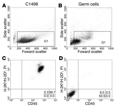
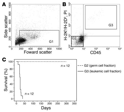
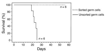
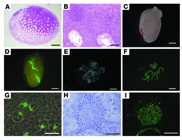
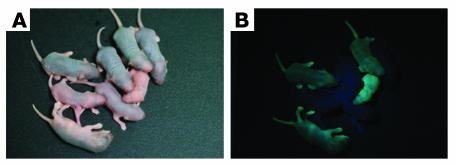
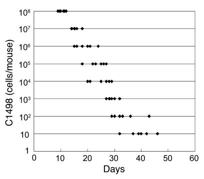
More than 70% of patients survive childhood leukemia, but chemotherapy and radiation therapy cause irreversible impairment of spermatogenesis. Although autotransplantation of germ cells holds promise for restoring fertility, contamination by leukemic cells may induce relapse. In this study, we isolated germ cells from leukemic mice by FACS sorting. The cell population in the high forward-scatter and low side-scatter regions of dissociated testicular cells from leukemic mice were analyzed by staining for MHC class I heavy chain (H-2K/H-2D) and for CD45. Cells that did not stain positively for H-2K/H-2D and CD45 were sorted as the germ cell-enriched fraction. The sorted germ cell-enriched fractions were transplanted into the testes of recipient mice exposed to alkylating agents. Transplanted germ cells colonized, and recipient mice survived. Normal progeny were produced by intracytoplasmic injection of sperm obtained from recipient testes. When unsorted germ cells from leukemic mice were transplanted into recipient testes, all recipient mice developed leukemia. The successful birth of offspring from recipient mice without transmission of leukemia to the recipients indicates the potential of autotransplantation of germ cells sorted by FACS to treat infertility secondary to anticancer treatment for childhood leukemia.
Figures

Similar articles
-
Isolation of germ cells from leukemia and lymphoma cells in a human in vitro model: potential clinical application for restoring human fertility after anticancer therapy.Cancer Res. 2006 Dec 1;66(23):11166-71. doi: 10.1158/0008-5472.CAN-06-2326. Cancer Res. 2006. PMID: 17145860
-
Restoration of fertility by germ cell transplantation requires effective recipient preparation.Biol Reprod. 2003 Aug;69(2):412-20. doi: 10.1095/biolreprod.103.016519. Epub 2003 Apr 2. Biol Reprod. 2003. PMID: 12672656
-
Stem cell based therapeutical approach of male infertility by teratocarcinoma derived germ cells.Hum Mol Genet. 2004 Jul 15;13(14):1451-60. doi: 10.1093/hmg/ddh166. Epub 2004 May 26. Hum Mol Genet. 2004. PMID: 15163638
-
Cryopreservation and transplantation of spermatogonia and testicular tissue for preservation of male fertility.J Natl Cancer Inst Monogr. 2005;(34):51-6. doi: 10.1093/jncimonographs/lgi029. J Natl Cancer Inst Monogr. 2005. PMID: 15784824 Review.
-
Germ cell transplantation and testis tissue xenografting in domestic animals.Anim Reprod Sci. 2005 Oct;89(1-4):137-45. doi: 10.1016/j.anireprosci.2005.06.020. Anim Reprod Sci. 2005. PMID: 16055282 Review.
Cited by
-
Alteration of spermatogenesis following spermatogonial stem cells transplantation in testicular torsion-detorsion mice.J Assist Reprod Genet. 2016 Jun;33(6):771-81. doi: 10.1007/s10815-016-0708-2. Epub 2016 Apr 6. J Assist Reprod Genet. 2016. PMID: 27052833 Free PMC article.
-
Spermatogonial stem cells: What does the future hold?Facts Views Vis Obgyn. 2011;3(1):36-40. Facts Views Vis Obgyn. 2011. PMID: 24753846 Free PMC article.
-
Characterization of human spermatogonial stem cell markers in fetal, pediatric, and adult testicular tissues.Reproduction. 2014 Oct;148(4):417-27. doi: 10.1530/REP-14-0123. Epub 2014 Jul 16. Reproduction. 2014. PMID: 25030892 Free PMC article.
-
Long-term health in recipients of transplanted in vitro propagated spermatogonial stem cells.Hum Reprod. 2018 Jan 1;33(1):81-90. doi: 10.1093/humrep/dex348. Hum Reprod. 2018. PMID: 29165614 Free PMC article.
-
Fertility preservation for boys with cancer.Reprod Med Biol. 2010 Aug 7;9(4):179-184. doi: 10.1007/s12522-010-0061-6. eCollection 2010 Dec. Reprod Med Biol. 2010. PMID: 29699341 Free PMC article. Review.
References
-
- U.S. Cancer Statistics Working Group. 2004. United States cancer statistics: 2000 incidence and mortality — updated and enhanced. Department of Health and Human Services, Centers for Disease Control and Prevention, and National Cancer Institute. Atlanta, Georgia, USA. http://apps.nccd.cdc.gov/uscs/index.asp?Year=2000.
-
- Jemal A, et al. Annual report to the nation on the status of cancer, 1975-2001, with a special feature regarding survival. Cancer. 2004;101:3–27. - PubMed
-
- Humpl T, Schramm P, Gutjahr P. Male fertility in long-term survivors of childhood ALL. Arch. Androl. 1999;43:123–129. - PubMed
-
- Thomson AB, et al. Semen quality and spermatozoal DNA integrity in survivors of childhood cancer: a case-control study. Lancet. 2002;360:361–367. - PubMed
-
- Bauld C, Anderson V, Arnold J. Psychosocial aspects of adolescent cancer survival. J. Paediatr. Child Health. 1998;34:120–126. - PubMed
MeSH terms
Substances
LinkOut - more resources
Full Text Sources
Other Literature Sources
Research Materials
Miscellaneous
