

A novel targeted therapy of Leydig and granulosa cell tumors through the luteinizing hormone receptor using a hecate-chorionic gonadotropin beta conjugate in transgenic mice
- PMID: 15967102
- PMCID: PMC1501163
- DOI: 10.1593/neo.04751
A novel targeted therapy of Leydig and granulosa cell tumors through the luteinizing hormone receptor using a hecate-chorionic gonadotropin beta conjugate in transgenic mice
Abstract
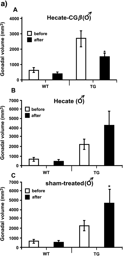
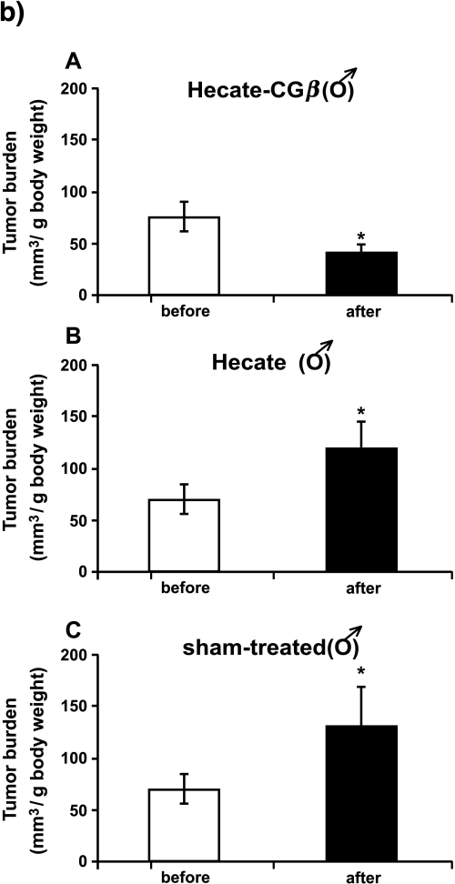
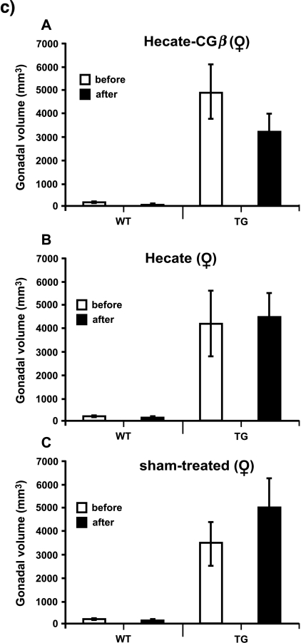
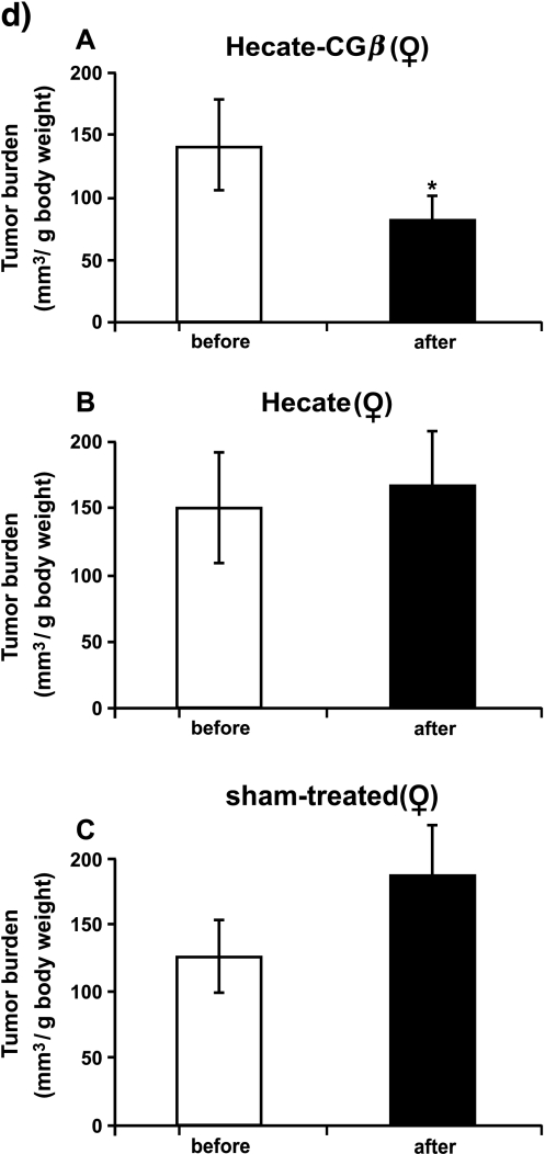
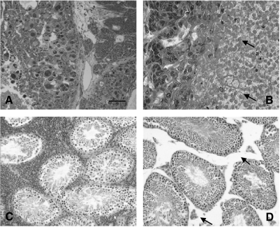
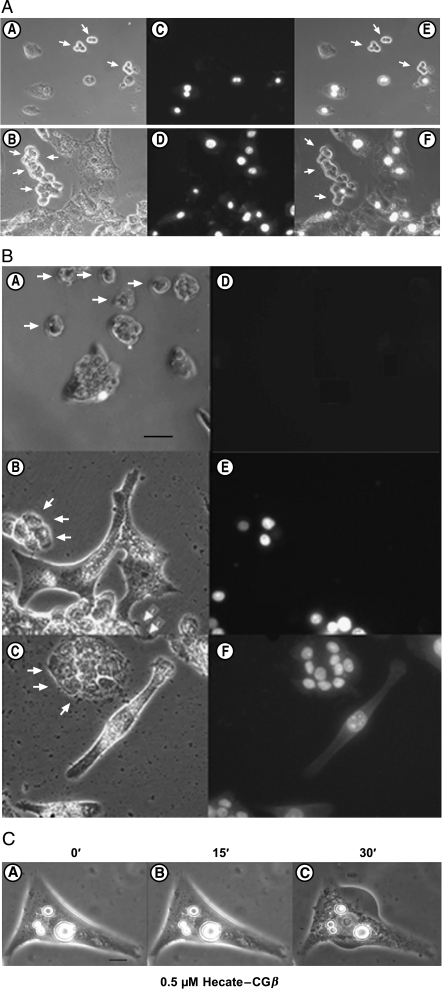
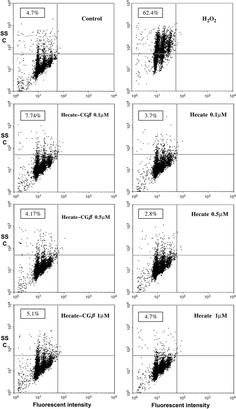
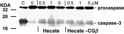
We investigated the antitumoral efficacy, endocrine consequences, and molecular mechanisms underlying cell death induced by the Hecate-chorionic gonadotropin (CG)beta conjugate, a fusion protein of a 23-amino acid lytic peptide Hecate with a 15-amino acid (81-95) fragment of the human CGbeta chain. Transgenic (TG) mice expressing the inhibin alpha-subunit promoter (inhalpha)/Simian Virus 40 T-antigen (Tag) transgene, developing luteinizing hormone (LH) receptor (R) expressing Leydig and granulosa cell tumors, and wild-type control littermates were treated either with vehicle, Hecate, or Hecate-CGbeta conjugate for 3 weeks. Hecate-CGbeta conjugate treatment reduced the testicular and ovarian tumor burden (P < .05), whereas a concomitant increase (testis; P < .05) or no change (ovary) in tumor volumes occured with Hectate treatment. A drop in serum progesterone, produced by the tumors, and an increase in LH levels occured in Hecate-CGbeta treated mice, in comparison with vehicle and Hecate groups, providing further support for the positive treatment response. Hecate-CGbeta conjugate induced a rapid and cell-specific membrane permeabilization of LHR-expressing cells in vitro, suggesting a necrotic mode of cell death without activation of apoptosis. These results prove the principle that the Hecate-CGbeta conjugate provides a novel specific lead into gonadal somatic cell cancer therapy by targeted destruction of LHR-expressing tumor cells.
Figures


Similar articles
-
Targeted therapy for adrenocortical tumors in transgenic mice through their LH receptor by Hecate-human chorionic gonadotropin beta conjugate.Endocr Relat Cancer. 2008 Jun;15(2):635-48. doi: 10.1677/ERC-08-0015. Endocr Relat Cancer. 2008. PMID: 18509010
-
Hecate-CGbeta conjugate and gonadotropin suppression shows two distinct mechanisms of action in the treatment of adrenocortical tumors in transgenic mice expressing Simian Virus 40 T antigen under inhibin-alpha promoter.Endocr Relat Cancer. 2009 Jun;16(2):549-64. doi: 10.1677/ERC-08-0232. Epub 2009 Mar 4. Endocr Relat Cancer. 2009. PMID: 19261682
-
A novel approach of targeted ablation of mammary carcinoma cells through luteinizing hormone receptors using Hecate-CGbeta conjugate.Breast Cancer Res Treat. 2003 May;79(1):1-10. doi: 10.1023/a:1023351819956. Breast Cancer Res Treat. 2003. PMID: 12779076
-
Use of hecate-chorionic gonadotropin beta conjugate in therapy of lutenizing hormone receptor expressing gonadal somatic cell tumors.Mol Cell Endocrinol. 2007 Apr 15;269(1-2):17-25. doi: 10.1016/j.mce.2006.11.016. Epub 2007 Feb 12. Mol Cell Endocrinol. 2007. PMID: 17363137 Review.
-
Transgenic mice expressing inhibin α-subunit promoter (inhα)/Simian Virus 40 T-antigen (Tag) transgene as a model for the therapy of granulosa cell-derived ovarian cancer.Reprod Biol. 2014 Mar;14(1):25-31. doi: 10.1016/j.repbio.2013.11.005. Epub 2013 Dec 11. Reprod Biol. 2014. PMID: 24607252 Review.
Cited by
-
C2AB: a molecular glue for lipid vesicles with a negatively charged surface.Langmuir. 2009 Jul 7;25(13):7177-80. doi: 10.1021/la901676e. Langmuir. 2009. PMID: 19563216 Free PMC article.
-
Human chorionic gonadotropin and its relation to grade, stage and patient survival in ovarian cancer.BMC Cancer. 2012 Jan 3;12:2. doi: 10.1186/1471-2407-12-2. BMC Cancer. 2012. PMID: 22214378 Free PMC article.
-
A review of the past, present, and future directions of neoplasia.Neoplasia. 2005 Dec;7(12):1039-46. doi: 10.1593/neo.05793. Neoplasia. 2005. PMID: 16354585 Free PMC article. Review. No abstract available.
-
Online monitoring of metabolism and morphology of peptide-treated neuroblastoma cancer cells and keratinocytes.J Bioenerg Biomembr. 2011 Jun;43(3):275-85. doi: 10.1007/s10863-011-9350-y. Epub 2011 Jun 4. J Bioenerg Biomembr. 2011. PMID: 21643697
-
GnRH antagonist treatment of malignant adrenocortical tumors.Endocr Relat Cancer. 2019 Jan 1;26(1):103-117. doi: 10.1530/ERC-17-0399. Endocr Relat Cancer. 2019. PMID: 30400009 Free PMC article.
References
-
- Freeman DA. Steroid hormone-producing tumors in man. Endocr Rev. 1986;7:204–220. - PubMed
-
- Kinkade S. Testicular cancer. Am Fam Physician. 1999;59:2539–2544. 2549–2550. - PubMed
-
- Crayford TJ, Campbell S, Bourne TH, Rawson HJ, Collins WP. Benign ovarian cysts and ovarian cancer: a cohort study with implications for screening. Lancet. 2000;355:1060–1063. - PubMed
-
- Goff BA, Mandel L, Muntz HG, Melancon CH. Ovarian carcinoma diagnosis. Cancer. 2000;89:2068–2075. - PubMed
-
- Malmstrom H, Hogberg T, Risberg B, Simonsen E. Granulosa cell tumors of the ovary: prognostic factors and outcome. Gynecol Oncol. 1994;52:50–55. - PubMed
Publication types
MeSH terms
Substances
LinkOut - more resources
Full Text Sources
Other Literature Sources
Molecular Biology Databases
Research Materials
Miscellaneous