

[18F]FDG uptake and PCNA, Glut-1, and Hexokinase-II expressions in cancers and inflammatory lesions of the lung
- PMID: 15967114
- PMCID: PMC1501150
- DOI: 10.1593/neo.04577
[18F]FDG uptake and PCNA, Glut-1, and Hexokinase-II expressions in cancers and inflammatory lesions of the lung
Abstract
Purpose: The aim of this study was to evaluate the relationships among [18F]fluorodeoxyglucose ([18F]-FDG) uptake, Glut-1 and HK-II expressions, and grade of inflammation in resected lung lesions.
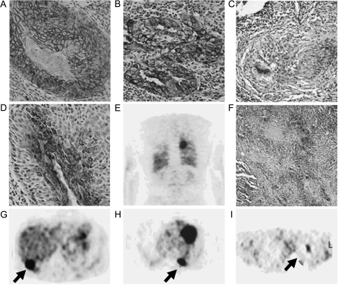
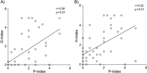
Materials and methods: Sixty patients had undergone preoperative 18F-FDG-PET imaging and thoracotomy. For semiquantitative analysis of 18F-FDG uptake, partial volume effect corrected maximum standardized uptake values (pSUVs) were calculated. Immunohistochemical staining was performed in resected specimens using anti-Glut-1, anti-HK-II, and anti-proliferative cellular nuclear antigen (PCNA) antibodies, and immunoreactivities were scored as G-, H-, and P-indexes on a five-point scale (0: 0%; 1: 20%, 2: 40%; 3: 60%; 4: 80%, and 5: 100% percentages of strongly immunoreactive cells).Grade of inflammation was also evaluated.
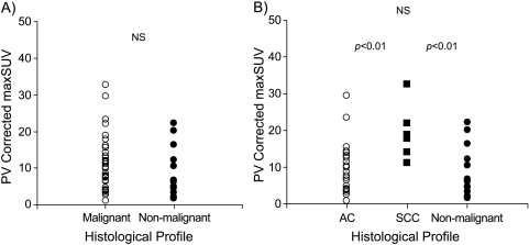
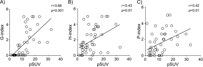
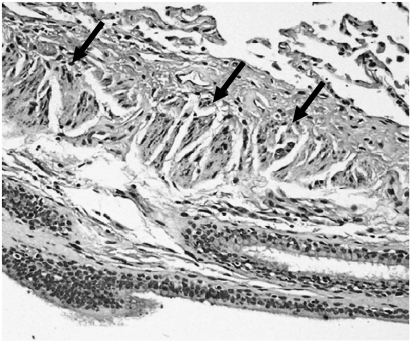
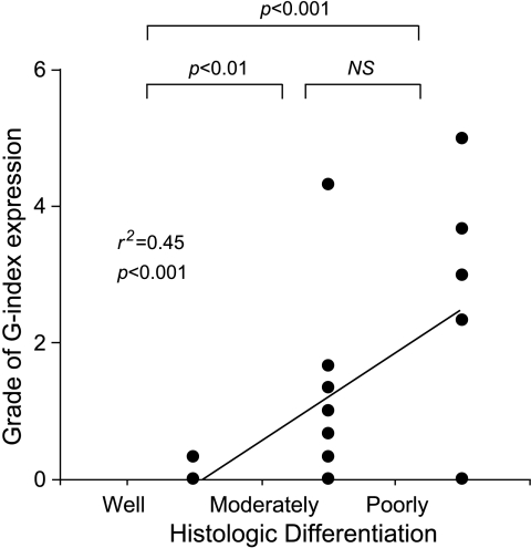
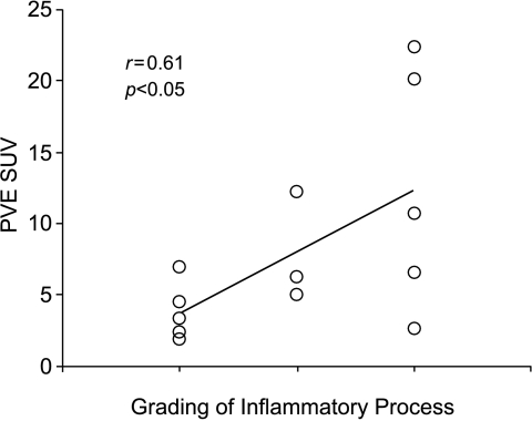
Results: The malignant lesions had higher pSUV and higher G- and H- than nonmalignant lesions. pSUVs correlated with the G- (p < .001), H- (p < .01), and P-indexes (p < .01) in malignant lesions. In adenocarcinomas, cancers with lower differentiation showed higher expression of Glut-1 and HK-II than those with higher differentiation. A positive linear regression was observed between pSUVs and the grading of inflammation in nonmalignant lesions (p < .05).
Conclusions: Our study indicates that 18F-FDG uptake in lung cancer correlates well with the Glut-1, HK-II, and PCNA expression. For nonmalignant lesions, the presence of a higher inflammatory process correlated with 18F-FDG uptake.
Figures
Similar articles
-
Correlation of Glut-1 glucose transporter expression with.Eur J Nucl Med. 2000 Dec;27(12):1778-85. Eur J Nucl Med. 2000. PMID: 11189940
-
Relationship between glucose transporter, hexokinase and FDG-PET in esophageal cancer.Hepatogastroenterology. 2005 Mar-Apr;52(62):486-90. Hepatogastroenterology. 2005. PMID: 15816463
-
Relationship between retention index in dual-phase (18)F-FDG PET, and hexokinase-II and glucose transporter-1 expression in pancreatic cancer.J Nucl Med. 2002 Feb;43(2):173-80. J Nucl Med. 2002. PMID: 11850481
-
FDG uptake, tumour characteristics and response to therapy: a review.Nucl Med Commun. 1998 Feb;19(2):97-105. doi: 10.1097/00006231-199802000-00002. Nucl Med Commun. 1998. PMID: 9548192 Review.
-
The rate-limiting step for tumor [18F]fluoro-2-deoxy-D-glucose (FDG) incorporation.Nucl Med Biol. 2001 Jan;28(1):1-4. doi: 10.1016/s0969-8051(00)00177-3. Nucl Med Biol. 2001. PMID: 11182558 Review. No abstract available.
Cited by
-
PET/CT imaging of Mycobacterium tuberculosis infection.Clin Transl Imaging. 2016;4:131-144. doi: 10.1007/s40336-016-0164-0. Epub 2016 Mar 7. Clin Transl Imaging. 2016. PMID: 27077068 Free PMC article. Review.
-
Overview of positron emission tomography in functional imaging of the lungs for diffuse lung diseases.Br J Radiol. 2022 Apr 1;95(1132):20210824. doi: 10.1259/bjr.20210824. Epub 2021 Nov 9. Br J Radiol. 2022. PMID: 34752146 Free PMC article. Review.
-
(18)F-FDG PET analysis of schwannoma: increase of SUVmax in the delayed scan is correlated with elevated VEGF/VPF expression in the tumors.Skeletal Radiol. 2009 Mar;38(3):261-6. doi: 10.1007/s00256-008-0612-7. Epub 2008 Dec 17. Skeletal Radiol. 2009. PMID: 19089420
-
Whole-tumor perfusion CT parameters and glucose metabolism measurements in head and neck squamous cell carcinomas: a pilot study using combined positron-emission tomography/CT imaging.AJNR Am J Neuroradiol. 2008 Aug;29(7):1376-81. doi: 10.3174/ajnr.A1111. Epub 2008 May 15. AJNR Am J Neuroradiol. 2008. PMID: 18483187 Free PMC article.
-
Specific biomarkers of receptors, pathways of inhibition and targeted therapies: clinical applications.Br J Radiol. 2011 Dec;84 Spec No 2(Spec Iss 2):S179-95. doi: 10.1259/bjr/76389842. Br J Radiol. 2011. PMID: 22433828 Free PMC article. Review.
References
-
- Jemal A, Tiwari RC, Murray T, Ghafoor A, Samuels A, Ward E, Feuer EJ, Thun MJ. Cancer statistics, 2004. CA Cancer J Clin. 2004;54:8–29. - PubMed
-
- Editorial Board of the Cancer Statistics in Japan, author; Foundation for Promotion on Cancer Research, editor. Cancer Statistics in Japan 2003. Tokyo: Foundation for Promotion of Cancer Research (FPCR); 2003. pp. 1–77.
-
- Warburg O, Posener K, Negelein E. The metabolism of the carcinoma cell. In: Warburg O, editor. The Mechanism of Tumors. New York, NY: Richard R. Smith, Inc.; 1931. pp. 129–169.
-
- Weber G. Enzymology of cancer cells. N Engl J Med. 1977;296:486–493. - PubMed
-
- Merrall NW, Plevin R, Gould GW. Growth factors, mitogens, oncogenes and the regulation of glucose transport. Cell Signal. 1993;5:667–675. - PubMed
MeSH terms
Substances
LinkOut - more resources
Full Text Sources
Medical
Miscellaneous