Comparative Study
. 2006 May;199(1):56-66.
doi: 10.1016/j.expneurol.2005.05.004. Epub 2005 Jun 20.

Intravenous infusion of immortalized human mesenchymal stem cells protects against injury in a cerebral ischemia model in adult rat

Affiliations
Comparative Study

Intravenous infusion of immortalized human mesenchymal stem cells protects against injury in a cerebral ischemia model in adult rat

T Honma et al. Exp Neurol. 2006 May.

Abstract

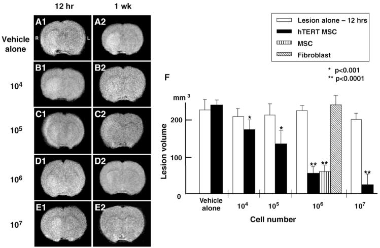
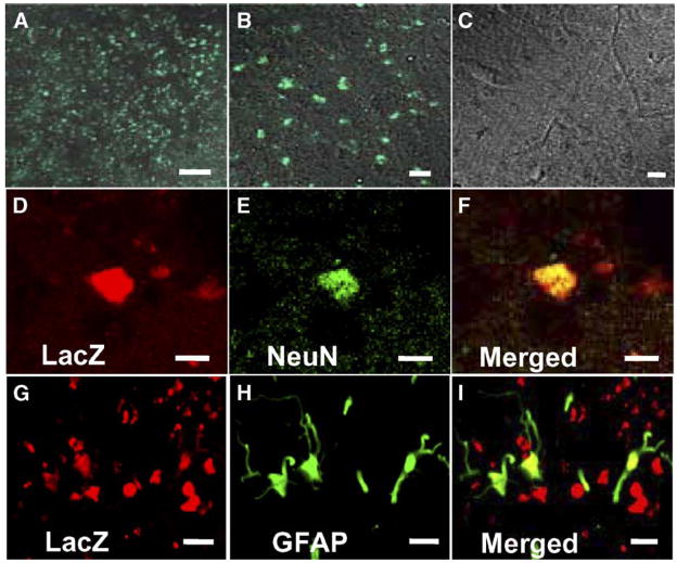
Intravenous infusion of bone marrow cells has demonstrated therapeutic efficacy in animal models of cerebral ischemia and spinal cord injury. We intravenously delivered human mesenchymal stem cells (SH2+, SH3+, CD34-, and CD45-) immortalized with a human-telomerase gene (hTERT-MSCs) and transfected with eGFP or LacZ into rats 12 h after induction of transient middle cerebral artery occlusion (MCAO), to study their potential therapeutic benefit. hTERT-MSCs were delivered at 12 h after lesion induction. Lesion size was assessed using MR imaging and spectroscopy, and histological methods. Functional outcome was assessed using the Morris water maze and a treadmill test. Intravenous delivery of hTERT-MSCs reduced lesion volume and the magnitude of the reduction and functional improvement was positively correlated with the number of cells injected. The reduction of lesion size could be assessed in vivo with MRI and MRS and was correlated with subsequent histological examination of the brain. This work demonstrates that highly purified hTERT-MSCs reduce cerebral infarction volume and improve functional outcome.


Figures

Fig. 1
Fig. 1
May–Giemsa staining of primary MSCs (A) and hTERT-MSCs (B) (scale bar = 20 μm). Flow cytometric analysis of the expression of surface antigens on primary MSCs (C) and hTERT-MSCs (D). The cells were immunolabeled with the FITC-conjugated monoclonal antibody specific for the indicated surface antigen. Dead cells were eliminated by forward and side scatter.
Fig. 2
Fig. 2
Evaluation of the ischemic lesion volume with MRI. T2-weighted images were obtained from experimental animals 12 h after MCAO and 1 week after intravenous delivery of varying concentrations of hTERT-MSCs. The left-hand column (panels A1–E1) shows single brain images obtained 12 h post injury. The right hand column (panels A2–E2) show images 1 week after treatment. Panel F indicates summary of the lesion volumes in each group.
Fig. 3
Fig. 3
Summary of the TTC-unstained lesion volumes in each group. Brain slices were stained with 2,3,5-triphenyltetrazolium chloride (TTC) to visualize the damaged lesions. A brain slice from the non-treated MCAO rats is shown for comparison (A). The TTC-stained brain slices from rats that were intravenously transplanted with 107 hTERT-MSCs 12 h after MCAO are shown in panel B. The sections were also counterstained with hematoxylin and eosin. Although the glial scar tissue and a large number of inflammatory cells were obvious in the lesion without cell transplantation (C), parenchymal brain tissue was greatly preserved in the treated group (D). Note that a small number of inflammatory cells were observed in the regions that may be heavily stressed by the ischemia. Panel E summarizes the TTC-unstained lesion volumes in each group. Scale bar = 3 mm (A, B), 20 μm (C, D).
Fig. 4
Fig. 4
Intravenously-administrated hTERT-MSCs accumulated in and around the ischemic lesions. Lower (A) and higher micrograph (B) demonstrating a large number of eGFP-positive cells in and around the lesion, but not in the non-treated rats (C). Confocal images show the differentiation of the transplanted hTERT-MSCs (LacZ in red; D, G) into neurons (NeuN in green; E) or astrocytes (GFAP in green; H). Panels F and I confirm the co-labeling of LacZ/NeuN or LacZ/GFAP in the cells, respectively. Scale bar = 250 μm (A), 10 mu;m (B, C), 5 mu;m (D–I).
Fig. 5
Fig. 5
Evaluation of cell therapy on stroke model rats by Magnetic Resonance Spectroscopy (MRS). NAA and lactate levels in the brain before and after cell transplantation were studied. Twelve hours (A, D, E) and 1 week (B, C, F, G) after MCAO induction, MRS analysis was carried out to access NAA and lactate levels in the lesioned (E–G) and non-lesioned hemispheres (D). With no cell transplantation, NAA signals were low and lactate signals were high 1 week after lesion induction (B, F). However, following intravenous delivery of 107 hTERT-MSCs, NAA signals were present and lactate signals were low (C, G). Scale bar = 5 mm (A–C).
Fig. 6
Fig. 6
The functional benefits following the intravenous administration of hTER-MSCs into the MCAO model rats were analyzed. The Treadmill test clearly demonstrates that the maximum speed at which the rats could run on a motor-driven treadmill was faster in the treated rats (A). The Morris water maze test also indicates that the learning and memory function was damaged in the MCAO model rats, but rescued and improved in the treated group (B). Days were plotted after MCAO.

Comment in

References

    1. Akiyama Y, Honmou O, Kato T, Uede T, Hashi K, Kocsis JD. Transplantation of clonal neural precursor cells derived from adult human brain establishes functional peripheral myelin in the rat spinal cord. Exp Neurol. 2001;167:27–39. - PubMed
    1. Akiyama Y, Radtke C, Honmou O, Kocsis JD. Remyelination of the spinal cord following intravenous delivery of bone marrow cells. Glia. 2002;39:229–236. - PMC - PubMed
    1. Alvarez-Dolado M, Pardal R, Garcia-Verdugo JM, Fike JR, Lee HO, Pfeffer K, Lois C, Morrison SJ, Alvarez-Buylla A. Fusion of bone-marrow-derived cells with Purkinje neurons, cardiomyocytes and hepatocytes. Nature. 2003 Oct. 30;425:968–973. - PubMed
    1. Azizi SA, Stokes D, Augelli BJ, DiGirolamo C, Prockop DJ. Engraftment and migration of human bone marrow stromal cells implanted in the brains of albino rats—Similarities to astrocyte grafts. Proc Natl Acad Sci U S A. 1998;95:3908–3913. - PMC - PubMed
    1. Barker PB, Gillard JH, van Zijl PC, Soher BJ, Hanley DF, Agildere AM, Oppenheimer SM, Bryan RN. Acute stroke: evaluation with serial proton MR spectroscopic imaging. Radiology. 1994;192(3):723–732. - PubMed

Publication types

MeSH terms