. 2005 Jun 20;169(6):941-52.
doi: 10.1083/jcb.200411155.

Conditional knockout of focal adhesion kinase in endothelial cells reveals its role in angiogenesis and vascular development in late embryogenesis

Affiliations

Conditional knockout of focal adhesion kinase in endothelial cells reveals its role in angiogenesis and vascular development in late embryogenesis

Tang-Long Shen et al. J Cell Biol. .

Abstract

Focal adhesion kinase (FAK) is a critical mediator of signal transduction by integrins and growth factor receptors in a variety of cells including endothelial cells (ECs). Here, we describe EC-specific knockout of FAK using a Cre-loxP approach. In contrast to the total FAK knockout, deletion of FAK specifically in ECs did not affect early embryonic development including normal vasculogenesis. However, in late embryogenesis, FAK deletion in the ECs led to defective angiogenesis in the embryos, yolk sac, and placenta, impaired vasculature and associated hemorrhage, edema, and developmental delay, and late embryonic lethal phenotype. Histologically, ECs and blood vessels in the mutant embryos present a disorganized, detached, and apoptotic appearance. Consistent with these phenotypes, deletion of FAK in ECs isolated from the floxed FAK mice led to reduced tubulogenesis, cell survival, proliferation, and migration in vitro. Together, these results strongly suggest a role of FAK in angiogenesis and vascular development due to its essential function in the regulation of multiple EC activities.


Figures
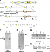
Figure 1.
Figure 1.
Generation of the floxed and total deletion FAK allele. (A) Schematic of mouse FAK protein, targeting vector, genomic and the loxP modified FAK loci. Large filled triangles represent loxP sites. The relevant restriction sites (A, ApaI; S, SacI; and B, BamHI), position of probe (thick line) for Southern blotting, and primers (P1, P2, and P3) for PCR genotyping are indicated. Crosses of the mice with targeted allele with EII-a Cre mice result in three possible outcomes at the FAK loci (flox, Δ and neoΔ). (B and C) Southern blotting analysis of the tail DNA from representative mice after digests with ApaI (B) or SacI (C). (D and E) Cell lysates were prepared from lung (D) or embryos (E) of mice with various genotypes as indicated. They were then analyzed by Western blotting using anti-FAK or anti-vinculin, as indicated.
Figure 2.
Figure 2.
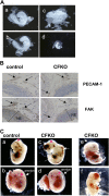
EC-specific FAK deletion in mice. (A) E8.5 embryos with +/+ (a), flox/Δ; Tie2Cre (CFKO) (b), flox/Δ (c), and Δ/Δ (d) genotypes. At this stage, CFKO embryos (c) are indistinguishable from control embryos (a and b), whereas FAK total KO embryos (d) showed a marked retardation in development and much reduced overall size. (B) Immunohistological analysis for PECAM-1 and FAK on adjacent brain sections of control and CFKO E9.5 embryos. Vascular ECs show positive staining with anti-PECAM-1 in both control and CFKO embryos (arrows). FAK is detected in the ECs of the control, but not CFKO, embryos (arrows). Neuroepithelium (NE, bracketed) are stained with FAK in both control and CFKO embryos. (C) Gross examination of whole embryos at E13.5 (a–d) or later (e and f), with (b and d) or without (a, c, e, and f) intact amnion and implantation sites. There are variable size hemorrhages (long arrows) in the CFKO embryos. Note the lack of vascular network in the head and amnion of the CFKO embryo when compared with the control littermates (red arrowheads). The amnion of CFKO embryos is thickened by edema (b and d, long arrows). The later embryos (e and f) are two examples of the representative range of abnormalities showing scattered hemorrhages and subcutaneous edema (e, long arrow), and smaller embryo with marked discoloration due to embryonic death and multifocal hemorrhages (f, long arrows).
Figure 3.
Figure 3.
Whole-mount staining of PECAM-1 of the embryos and yolk sacs. (A) E10.5 embryos. There is no difference of vascular structures at this stage between the control and CFKO embryos. (B) E13.5 embryos. Typical defects in the vascular network observed in the CFKO embryos are shown. There is lack of the superficial vascular network in the head region (long arrows), no clearly defined visceral outlines, and no clear definition of the axial skeleton at the coccigeal level (corresponding part marked by short arrow for the control embryo) when compared with the control embryo. (C) Characteristic decrease of superficial vasculature with absence of small vessel branch formation in the yolk sacs of CFKO embryos.
Figure 4.
Figure 4.
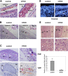
Vascular lesions and associated defects of CFKO embryos and placentas. (A) Histopathological sections of skin from the dorsum of control and CFKO embryos. There is marked expansion of the dermis and subcutaneous tissue of the CFKO embryos by hemorrhage and edema. RBC, red blood cells. (B) Histopathological section of a median plane at the level of the thoracolumbar region of control and CFKO embryos. Distinctive expansion of the subcutaneous tissue in the CFKO embryos due to edema and engorged capillaries with perivascular hemorrhages; the brown fat (BF) pads are smaller with pyknotic nuclei and scant eosinophilic cytoplasm. The muscle mass (MM) is reduced significantly (bracketed). Insets show RBC inside the EC-lined vessels in control embryos, but both inside and outside of the capillaries in CFKO embryos. (C) Hematoxylin and eosin staining and immunohistochemical staining with anti-vWF show the normal capillaries with RBC in the control embryos and collapsed vessels lined by defective ECs in the CFKO embryos. Some of the ECs are pyknotic with karyorrhectic debris (red arrowheads), others are swollen and rounded (arrows), and a few have sloughed into the lumen of the vessel. The collapsed vessels are outlined with lumina marked by asterisks. (D) Hoechst staining of control and CFKO embryos. Presence of apoptotic ECs in the CFKO (but not control) embryos are marked by the long arrow. (E) Histopathological sections from E13.5 embryos implantation site. Typical examples of decreased thickness of the labyrinth area in the CFKO embryos compared with the controls (top panels). The arterial canals and branches on the CFKO labyrinth have collapsed vessel outlines and the lumina are devoid of nucleated fetal RBCs (bottom panels and graph). D, decidua; L, labyrinth layer; T, trophoblast.
Figure 5.
Figure 5.
Defective tubulogenesis of the isolated FAK −/− primary ECs. (A) ECs isolated from homozygous floxed FAK mice (flox/flox) were infected with increasing amount of recombinant adenoviruses encoding Cre (Ad-Cre) or a control insert (Ad-lacZ), as indicated. Cell lysates were analyzed by Western blotting with anti-FAK or anti-vinculin (top two panels). Genomic DNA was analyzed by PCR (bottom two panels). (B–D) Primary ECs from floxed FAK mice and infected with Ad-Cre or Ad-lacZ were cultured on Matrigel as described in Materials and methods. Images of representative fields are shown in B. The length of the tubules (C) and branch points (D) were quantified from three independent experiments and shown as the relative ratio of the value + standard error. *, P = 0.023 and **, P = 0.017 in comparison to value from Ad-lacZ–induced cells.
Figure 6.
Figure 6.
Increased apoptosis, and reduced proliferation and migration of the isolated FAK −/− primary ECs. Primary ECs from floxed FAK mice and infected with Ad-lacZ or Ad-Cre were measured for apoptosis using TUNEL assay (A), proliferation by BrdU incorporation assay (B), and migration in response to VEGF or FN by Boyden chamber assay (C) and wound closure assay (D), as described in Materials and methods. The mean + standard error from at least three experiments is shown. *, P = 0.014 and **, P < 0.001 in comparison to value from Ad-lacZ–infected cells.
Figure 7.
Figure 7.
Differential requirement of FAK kinase activity for VEGF-stimulated EC migration. (A–C) Primary ECs from floxed FAK mice were infected with Ad-Cre to delete endogenous FAK followed by infection of Ad-FAK, Ad-KD, or the control Ad-GFP as indicated. Aliquots of lysates were analyzed by Western blotting using anti-FAK, anti-pY397, or anti-vinculin, as indicated (A). The infected cells were subjected to wound closure assay in response to VEGF (B) or FN (C), as described in Materials and methods. The mean + standard error from at least three experiments is shown. *, P < 0.005; **, P = 0.448; and ***, P = 0.012 in comparison to value from Ad-GFP–infected cells. (D and E) MEFs were isolated from floxed FAK mice, and then infected with Ad-lacZ or Ad-Cre (D) or Ad-Cre followed by Ad-FAK, Ad-KD, or the control Ad-GFP (E), as indicated. The infected cells were then subjected to wound closure assays in response to VEGF (D as indicated) or FN (D as indicated and E). The mean + standard error from at least three experiments is shown. *, P < 0.01 and **, P = 0.17 in comparison to value from (D) Ad-lacZ– or (E) Ad-GFP–infected cells.
Figure 8.
Figure 8.
Effects of FAK deletion on downstream targets in the isolated primary ECs. (A) Cell lysates of primary ECs from floxed FAK mice and infected with Ad-lacZ or Ad-Cre were analyzed by Western blotting with various antibodies as indicated. (B) Immunofluorescent staining of the above ECs (see A) with anti-phosphotyrosine antibody PY20 (top panels) or anti-vinculin (bottom panels). Focal adhesions are marked by arrows.

References

    1. Burridge, K., C.E. Turner, and L.H. Romer. 1992. Tyrosine phosphorylation of paxillin and pp125FAK accompanies cell adhesion to extracellular matrix: a role in cytoskeletal assembly. J. Cell Biol. 119:893–903. - PMC - PubMed
    1. Cary, L.A., J.F. Chang, and J.L. Guan. 1996. Stimulation of cell migration by overexpression of focal adhesion kinase and its association with Src and Fyn. J. Cell Sci. 109:1787–1794. - PubMed
    1. Cary, L.A., D.C. Han, T.R. Polte, S.K. Hanks, and J.L. Guan. 1998. Identification of p130Cas as a mediator of focal adhesion kinase-promoted cell migration. J. Cell Biol. 140:211–221. - PMC - PubMed
    1. Cattelino, A., S. Liebner, R. Gallini, A. Zanetti, G. Balconi, A. Corsi, P. Bianco, H. Wolburg, R. Moore, B. Oreda, et al. 2003. The conditional inactivation of the beta-catenin gene in endothelial cells causes a defective vascular pattern and increased vascular fragility. J. Cell Biol. 162:1111–1122. - PMC - PubMed
    1. Chen, J., F. Young, A. Bottaro, V. Stewart, R.K. Smith, and F.W. Alt. 1993. Mutations of the intronic IgH enhancer and its flanking sequences differentially affect accessibility of the JH locus. EMBO J. 12:4635–4645. - PMC - PubMed

Publication types

MeSH terms