. 2005 Jun 20;169(6):977-85.
doi: 10.1083/jcb.200411153.

Intracellular collagen degradation mediated by uPARAP/Endo180 is a major pathway of extracellular matrix turnover during malignancy

Affiliations

Intracellular collagen degradation mediated by uPARAP/Endo180 is a major pathway of extracellular matrix turnover during malignancy

Alejandro C Curino et al. J Cell Biol. .

Abstract

We recently reported that uPARAP/Endo180 can mediate the cellular uptake and lysosomal degradation of collagen by cultured fibroblasts. Here, we show that uPARAP/Endo180 has a key role in the degradation of collagen during mammary carcinoma progression. In the normal murine mammary gland, uPARAP/Endo180 is widely expressed in periductal fibroblast-like mesenchymal cells that line mammary epithelial cells. This pattern of uPARAP/Endo180 expression is preserved during polyomavirus middle T-induced mammary carcinogenesis, with strong uPARAP/Endo180 expression by mesenchymal cells embedded within the collagenous stroma surrounding nests of uPARAP/Endo180-negative tumor cells. Genetic ablation of uPARAP/Endo180 impaired collagen turnover that is critical to tumor expansion, as evidenced by the abrogation of cellular collagen uptake, tumor fibrosis, and blunted tumor growth. These studies identify uPARAP/Endo180 as a key mediator of collagen turnover in a pathophysiological context.


Figures

Figure 1.
Figure 1.
uPARAP/Endo180 is expressed in fibroblast-like stromal cells of the normal murine mammary gland and mammary tumors. (A) Immunohistochemical staining of normal virgin mammary gland showing expression of uPARAP/Endo180 in fibroblast-like mesenchymal cells lining the periphery of mammary ducts (arrow) and fibroblast-like cells embedded in adipose tissue (arrowheads), but not in mammary epithelial cells (curved arrow). (B) Mammary intraepithelial neoplasia in a 5-wk-old PymT female mouse showing periductal expression of uPARAP/Endo180 in fibroblast-like mesenchymal cells (arrows), but absence of uPARAP/Endo180 expression in mammary tumor cells (stars). (C) Advanced mammary carcinoma in a 13-wk-old PymT mouse showing uPARAP/Endo180 expression in fibroblast-like cells in connective tissue streaks transecting tumors (arrows), and absence of uPARAP/Endo180 expression in mammary tumor cells (star). (D) Absence of immunoreactivity in a tumor from a 13-wk-old PymT/uPARAP/Endo180−/− mouse stained in parallel demonstrates the specificity of the immunostaining procedure for uPARAP/Endo180. Brightfield (E) and darkfield (F) images of in situ hybridization using a uPARAP antisense probe of a mammary tumor from a 15-wk-old mouse, demonstrating the identical distribution of uPARAP/Endo180 mRNA and antigen, with uPARAP/Endo180 mRNA in stromal cells (arrowheads) adjacent to nests of uPARAP/Endo180 mRNA-negative tumor cells (stars). Bars, 50 μm.
Figure 2.
Figure 2.
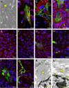
uPARAP/Endo180 mediates cellular collagen uptake in mammary tumors. (A) Phase-contrast micrograph of explanted uPARAP/Endo180+ mammary tumors showing spontaneous organization of tightly clustered sheets of small epithelial tumor cells (star) surrounded by larger mesenchymal cells with a typical fibroblast morphology. (B and C) Representative confocal fluorescence images of explants of mammary tumors from uPARAP/Endo180+ (B) and littermate uPARAP/Endo180−/− (C) mice 8 h after the addition of Oregon green–conjugated collagen fibrils to the explant medium. Explants were stained with LysoTracker (red) for the visualization of lysosomes and DAPI (blue) for the visualization of nuclei. uPARAP/Endo180+ tumor explants display the uptake of fluorescence-labeled collagen (green) into vesicles of large mesenchymal, fibroblast-like stromal cells located adjacent to mammary tumor cell clusters. Targeting of collagen to lysosomes is shown by the colocalization of green and red fluorescence (B, yellow) in some perinuclear vesicles. In sharp contrast to uPARAP/Endo180+ explants, collagen deposits in extracellular fiberlike structures in uPARAP/Endo180−/− mammary tumor explants. (D) Confocal fluorescence images after labeling of tumor cells with cytokeratin antibodies (red) shows the absence of internalized collagen in tumor cells. (E–J) Time course analysis of collagen internalization in uPARAP/Endo180+ (E, G, and I) and uPARAP/Endo180−/− explants (F, H, and J) using Oregon green–conjugated collagen fibrils and LysoTracker for visualization of lysosomes. Diffuse localization of collagen with occasional fiberlike structures immediately after collagen addition (E and F). Increased cellular association after 2 h, with some collagen in intracellular vesicles in uPARAP/Endo180+ explants (G), but not in uPARAP/Endo180−/− explants (H). (I) Predominant localization of collagen in intracellular vesicles of stromal cells of uPARAP/Endo180+ explants at 4 h, frequently colocalizing with LysoTracker-positive vesicles with perinuclear locations, versus the location of collagen in extracellular fiberlike structures with an increasing degree of organization compared with those after 2 h in uPARAP/Endo180−/− explants (J). (K and L) Representative examples of high magnification transmission electron micrographs of mammary tumor stromal cells from 105-d-old mice showing intracellular collagen inclusion (star) that is adjacent to the nucleus of a uPARAP/Endo180+ fibroblast (K) and the exclusively extracellular localization of collagen (star) in uPARAP/Endo180−/− tumors (L). Arrows (L) show the position of the plasma membrane. Bars: (B–J) 40 μm; (K and L) 500 nm.
Figure 3.
Figure 3.
Collagen accumulation in uPARAP/Endo180-deficient mammary tumors. Representative examples of the appearance of uPARAP/Endo180+ tumors (A, C, E, G, and I) and uPARAP/Endo180−/− tumors (B, D, F, H, and J) after immunohistochemical staining for collagen type I (A and B), type IV (C and D), fibronectin (E and F), decorin, (G and H), and nidogen (I and J). uPARAP/Endo180−/− tumors display an obvious accumulation of collagen in the tumor stroma (arrowheads) surrounding nests of tumor cells (A–D, arrows), whereas no significant changes are apparent in fibronectin, nidogen, or decorin deposition (arrowheads) around tumor cell nests (E–J, arrows). (K) Histomorphometric quantitation of collagen I, collagen IV, fibronectin, nidogen, and decorin accumulation in uPARAP/Endo180+ (crosshatched bars) and uPARAP/Endo180−/− (shaded bars) tumors. (L) Northern blot analysis of Col1A1 and Col4A1 mRNA in uPARAP/Endo180+ (crosshatched bars) and uPARAP/Endo180−/− (shaded bars) tumors. Signal intensities were determined by PhosphorImage analysis and normalized to the GAPDH mRNA signal intensity. Error bars indicate SEM. (M) Gelatin zymography (top) of extracts of uPARAP/Endo180+ (lanes 1–3) and uPARAP/Endo180−/− tumors (lanes 4–6), and reverse zymography (bottom) showing amounts of zymographically active MMP-2, MMP-9, and TIMP-2. The position of lysis zones generated by MMP-2 and active MMP-9 and zones of inhibition of lysis by TIMP-2 are indicated at left. The positions of molecular mass markers (kD) are indicated at right. (N–U) Representative examples of in situ hybridization of MT1-MMP (N, O, R, and S) and MMP-13 (P, Q, T, and U) mRNA expression in uPARAP/Endo180−/− tumors (N–Q) and uPARAP/Endo180+ tumors (R–U) using 32P-labeled antisense probes and complementary sense probes. Specific hybridization signals in uPARAP/Endo180-sufficient and -deficient tumors are observed in stromal cells (arrowheads) surrounding nests of tumor cells (arrows). Six tumors were analyzed for each of the two MMPs. Bars: (A–J) 500 μm; (N–U) 100 μm. All p-values were determined by a two-tailed t test.
Figure 4.
Figure 4.
uPARAP/Endo180 promotes mammary tumor progression. Scatter plots of the cumulative tumor burdens of uPARAP/Endo180+ (open circles) and uPARAP/Endo180−/− (filled circles) mice at 95 (A) and 105 d of age (B), as determined by postmortem measurement of the weight of excised mammary tumors. Horizontal bars indicate mean values. P-values determined by a two-tailed t test and a Wilcoxon rank sum test are shown. N indicates the number of mice analyzed.

References

    1. Behrendt, N. 2004. The urokinase receptor (uPAR) and the uPAR-associated protein (uPARAP/Endo180): membrane proteins engaged in matrix turnover during tissue remodeling. Biol. Chem. 385:103–136. - PubMed
    1. Bernstein, E.F., Y.Q. Chen, J.B. Kopp, L. Fisher, D.B. Brown, P.J. Hahn, F.A. Robey, J. Lakkakorpi, and J. Uitto. 1996. Long-term sun exposure alters the collagen of the papillary dermis. Comparison of sun-protected and photoaged skin by northern analysis, immunohistochemical staining, and confocal laser scanning microscopy. J. Am. Acad. Dermatol. 34:209–218. - PubMed
    1. Blavier, L., A. Lazaryev, J. Groffen, N. Heisterkamp, Y.A. DeClerck, and V. Kaartinen. 2001. TGF-beta3-induced palatogenesis requires matrix metalloproteinases. Mol. Biol. Cell. 12:1457–1466. - PMC - PubMed
    1. Bugge, T.H., L.R. Lund, K.K. Kombrinck, B.S. Nielsen, K. Holmback, A.F. Drew, M.J. Flick, D.P. Witte, K. Danø, and J.L. Degen. 1998. Reduced metastasis of Polyoma virus middle T antigen-induced mammary cancer in plasminogen-deficient mice. Oncogene. 16:3097–3104. - PubMed
    1. Chambers, A.F., A.C. Groom, and I.C. MacDonald. 2002. Dissemination and growth of cancer cells in metastatic sites. Nat. Rev. Cancer. 2:563–572. - PubMed

Publication types

MeSH terms

Associated data