. 2005 Jun 28;102(26):9194-9.
doi: 10.1073/pnas.0503280102. Epub 2005 Jun 20.

Cell intrinsic alterations underlie hematopoietic stem cell aging

Affiliations

Cell intrinsic alterations underlie hematopoietic stem cell aging

Derrick J Rossi et al. Proc Natl Acad Sci U S A. .

Abstract

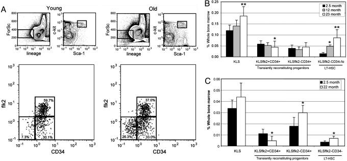
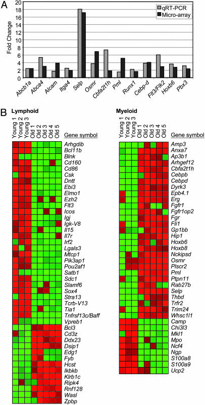
Loss of immune function and an increased incidence of myeloid leukemia are two of the most clinically significant consequences of aging of the hematopoietic system. To better understand the mechanisms underlying hematopoietic aging, we evaluated the cell intrinsic functional and molecular properties of highly purified long-term hematopoietic stem cells (LT-HSCs) from young and old mice. We found that LT-HSC aging was accompanied by cell autonomous changes, including increased stem cell self-renewal, differential capacity to generate committed myeloid and lymphoid progenitors, and diminished lymphoid potential. Expression profiling revealed that LT-HSC aging was accompanied by the systemic down-regulation of genes mediating lymphoid specification and function and up-regulation of genes involved in specifying myeloid fate and function. Moreover, LT-HSCs from old mice expressed elevated levels of many genes involved in leukemic transformation. These data support a model in which age-dependent alterations in gene expression at the stem cell level presage downstream developmental potential and thereby contribute to age-dependent immune decline, and perhaps also to the increased incidence of leukemia in the elderly.


Figures

Fig. 1.
Fig. 1.
Increased self-renewal of LT-HSCs as a consequence of age. (A) Representative FACS profiles of young and old BM cells showing HPCs and multipotent progenitor cells. (B) BM frequencies of KLS cell subsets from young (n = 16), middle-aged (n = 5), and old mice (n = 17). (C) BM frequencies of donor-derived KLS cell subsets after transplantation of BM from young or old donors into young recipients 4 months posttransplant (n = 4 young, n = 3 old). Recipient mice were transplanted with 5-fold more young cells to approximate stem cell equivalents. Statistically significant differences (P < 0.05) are indicated by *. ** in B indicate statistical significance to both of the other age groups.
Fig. 2.
Fig. 2.
Aging is associated with stem cell intrinsic alterations in lineage and progenitor potential. (A) Multilineage reconstitution (Left) and lineage potential (Right) from transplant experiments of LT-HSCs from young or old mice. Fifty KLSflk2-CD34- cells purified from young or old CD45.2 donors were transplanted into congenic (CD45.1) young recipients against 3 × 10⁵ competitor cells. Peripheral blood analysis was done at the indicated time points, and donor repopulating ability is presented as the percent of total white blood cells. Donor contribution to lymphoid and myeloid lineages is presented as the percent of B, myeloid, and T cells. (B) Aged BM microenvironment does not affect the lineage distribution of adoptively transferred LT-HSCs long term. Ninety LT-HSCs from young mice were transplanted into congenic young or old recipients along with 3 × 10⁵ competitor BM cells from old donors. Peripheral blood analysis was done at the indicated time points, and donor-derived contribution to B cells and myeloid cells is presented as the percent of T cell receptor (TCR) β-negative cells so as not to bias the B cell and myeloid cell data by negating the contribution of the involuted thymus in the old recipients. The contribution to T cells was looked at separately by gating on total donor-derived contribution to all lineages before assessing T cell contribution (Right). (C) BM frequencies of CMPs, GMPs, and MEPs from young (n = 10) and old mice (n = 9). (D) BM frequencies of CLP in young (n = 8), middle-aged (n = 5), and old mice (n = 7). (E) BM frequencies of donor-derived myeloid progenitors from either young or old donors into young recipient mice 4 months posttransplant (n = 4 young, n = 3 old). (F) BM frequencies of donor-derived CLPs from either young or old donors into young recipient mice 4 months posttransplant (young n = 9, old n = 4). Statistically significant differences are indicated by *; ** indicates statistical significance to both age groups.
Fig. 3.
Fig. 3.
Down-regulation of lymphoid genes and the up-regulation of myeloid genes in aged LT-HSCs. (A) Validation of microarray data by qRT-PCR. Fold change of expression of several age-regulated genes as determined by qRT-PCR (light gray) and our microarray analysis (dark gray) is shown. qRT-PCR fold change is presented as the average of three independent experiments. (B) Heat map showing the expression of all of the age-regulated genes identified as lymphoid-specific (Left) and myeloid-specific (Right) in aging LT-HSCs. Gene up-regulation in old LT-HSCs is presented in red and gene down-regulation is in green.

References

    1. Schlessinger, D. & Van Zant, G. (2001) Mech. Ageing Dev. 122, 1537-1553. - PubMed
    1. Morrison, S. J., Wandycz, A. M., Akashi, K., Globerson, A. & Weissman, I. L. (1996) Nat. Med. 2, 1011-1016. - PubMed
    1. Ikuta, K., Kina, T., MacNeil, I., Uchida, N., Peault, B., Chien, Y. H. & Weissman, I. L. (1990) Cell 62, 863-874. - PubMed
    1. Hardy, R. R., Wei, C. J. & Hayakawa, K. (2004) Immunol. Rev. 197, 60-74. - PubMed
    1. Albright, J. W. & Makinodan, T. (1976) J. Exp. Med. 144, 1204-1213. - PMC - PubMed

Publication types

MeSH terms