

The role of popliteal lymph nodes in differentiating rheumatoid arthritis from osteoarthritis by using CE 3D FSPGR MR imaging: relationship of the inflamed synovial volume
- PMID: 15968151
- PMCID: PMC2686419
- DOI: 10.3348/kjr.2005.6.2.117
The role of popliteal lymph nodes in differentiating rheumatoid arthritis from osteoarthritis by using CE 3D FSPGR MR imaging: relationship of the inflamed synovial volume
Abstract
Objective: We wanted to assess the role of the popliteal lymph nodes for differentiating rheumatoid arthritis (RA) from osteoarthritis (OA), and we also wanted to investigate the relationship between the popliteal lymph nodes and the inflamed synovial volume (ISV) by using contrast enhanced (CE), fat suppressed, three dimensional-fast spoiled gradient echo (3D-FSPGR) MR imaging.
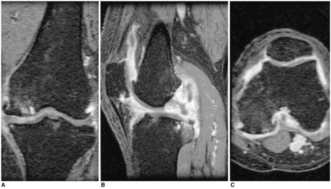
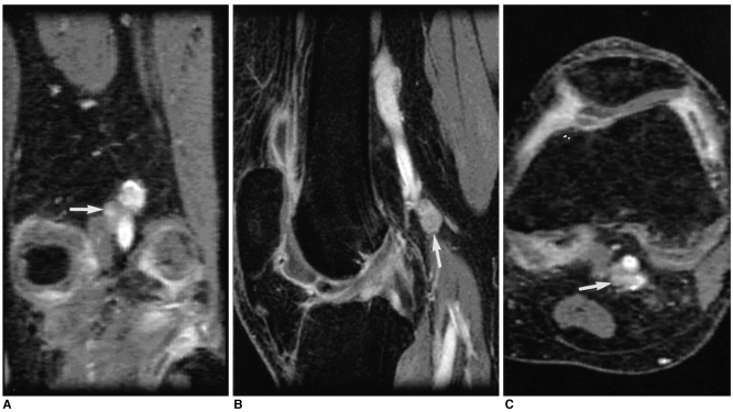
Materials and methods: Contrast enhanced 3D-FSGPR MR imaging of 94 knees (21 with RA and 73 with OA) was analyzed. A lymph node was defined as being 'observed' if it could be seen in at least two planes of the three orthogonal reformatted planes. The number of observed lymph nodes, the mean of the smallest dimension of each lymph node and the existence of central fatty change were recorded. The OA group was graded according to the ISV calculated by a segmentation method: grade I was < 20 cm(3); grade II ranged from 20 cm(3) to 40 cm(3); and grade III was > 40 cm(3). Statistical analysis of the number and the mean size of the popliteal lymph nodes among the four groups (the RA group and the grade I-III OA groups) was performed.
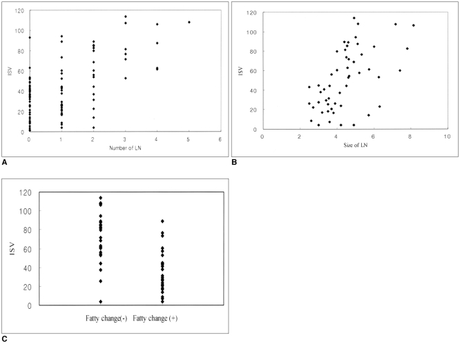
Results: The prevalence of the observed popliteal lymph nodes was significantly different between all the OA groups or between the grade III OA group and the RA group (p < 0.0001, 0.0001, respectively). The popliteal lymph node was observed in 32 out of 73 OA cases, whereas it was visible in all of the 21 RA cases. The number (mean +/- standard deviation) of lymph nodes in the grade I OA group, the grade II OA group, the grade III OA group and the RA group was 1.2 +/- 0.4, 1.2 +/- 0.4, 1.3 +/- 0.5, and 2.7 +/- 1.1, respectively. The mean size (mean +/- standard deviation) of the lymph nodes was 3.8 +/- 1.0 mm, 3.6 +/- 1.1 mm, 4.1 +/- 0.8 mm, and 5.4 +/- 1.3 mm, respectively. The incidence of central fatty changes was significantly lower in the RA group than in all the OA groups and the grade III OA group. When differentiating RA from OA, and when the differentiation was confined to the RA group and grade III OA group, respectively, the criteria of the number of lymph nodes, their size, their central fatty change and a combination of all these three criteria showed statistical significance (Az values for the former were 0.869, 0.847, 0.776, and 0.942; Az values for the latter were 0.855, 0.799, 0.712, and 0.916). The number and mean size of the lymph nodes correlated with the ISVs (r = 0.49, p < 0.001; 0.50, 0.001, respectively).
Conclusion: The number, size and central fatty changes in the popliteal lymph nodes observed on the MR images might serve as simple and useful markers in differentiating RA disease from OA disease. These markers would be particular helpful in cases of severe synovial enhancement where the ISVs of both RA and OA overlap. The number and mean size of the lymph nodes also correlated well with the ISV.
Figures
References
-
- Netter Frank H. The Ciba collection of medical illustrations. 3rd ed. Newjersey: Ciba-Geigy Corp; 1987. p. 121.
-
- William PL, Warwick R, Dyson M, Bannister LH. Gray's Anatomy. 37th ed. London: Churchill Livingstone; 1989. pp. 848–849.
-
- Moon HJ, Suh JS, Lee SH. Magnetic resonance appearance of normal popliteal lymph nodes: location and relationship of number, fatty change, and size of the lymph nodes with aging. J Korean Radiol Soc. 2002;47:665–671.
-
- Huh YM, Suh JS, Jeong EK, Lee SK, Lee JS, Choi BW, et al. Role of the inflamed synovial volume of the wrist in defining remission of rheumatoid arthritis with gadolinium-enhanced 3D-SPGR MR imaging. J Magn Reson Imaging. 1999;10:202–208. - PubMed
Publication types
MeSH terms
LinkOut - more resources
Full Text Sources
Other Literature Sources
Medical
