. 2005 Aug;77(2):242-51.
doi: 10.1086/432556. Epub 2005 Jun 20.

A mutation in SNAP29, coding for a SNARE protein involved in intracellular trafficking, causes a novel neurocutaneous syndrome characterized by cerebral dysgenesis, neuropathy, ichthyosis, and palmoplantar keratoderma

Affiliations

A mutation in SNAP29, coding for a SNARE protein involved in intracellular trafficking, causes a novel neurocutaneous syndrome characterized by cerebral dysgenesis, neuropathy, ichthyosis, and palmoplantar keratoderma

Eli Sprecher et al. Am J Hum Genet. 2005 Aug.

Abstract

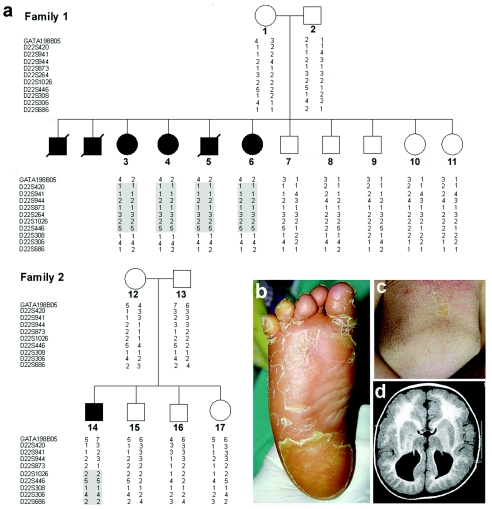
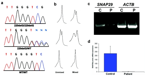
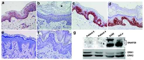
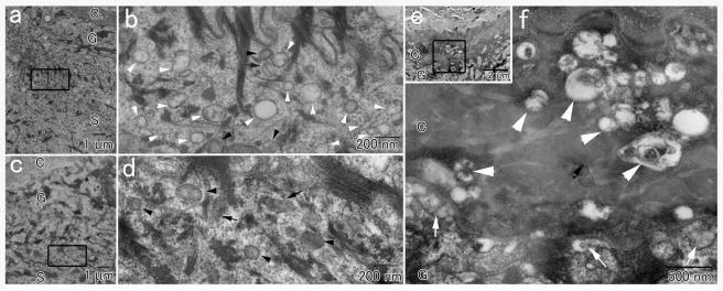
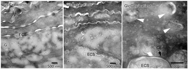
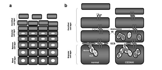
Neurocutaneous syndromes represent a vast, largely heterogeneous group of disorders characterized by neurological and dermatological manifestations, reflecting the common embryonic origin of epidermal and neural tissues. In the present report, we describe a novel neurocutaneous syndrome characterized by cerebral dysgenesis, neuropathy, ichthyosis, and keratoderma (CEDNIK syndrome). Using homozygosity mapping in two large families, we localized the disease gene to 22q11.2 and identified, in all patients, a 1-bp deletion in SNAP29, which codes for a SNARE protein involved in vesicle fusion. SNAP29 expression was decreased in the skin of the patients, resulting in abnormal maturation of lamellar granules and, as a consequence, in mislocation of epidermal lipids and proteases. These data underscore the importance of vesicle trafficking regulatory mechanisms for proper neuroectodermal differentiation.


Figures

Figure  1
Figure 1
Family trees, genotyping, and clinical spectrum of CEDNIK syndrome. a, Haplotype analysis in two families affected with CEDNIK syndrome, performed using polymorphic microsatellite markers on 22q11.2. Homozygous disease-associated haplotypes for each family are marked in gray. b, Severe thickening (keratoderma) of the plantar skin in patient 3. c, Lamellar ichthyosis over the abdominal surface in patient 14. d, Axial T1 (SE) weighted MR image demonstrating cortical dysplasia and pachygyria with polymicrogyria, as well as typical absence of corpus callosum, in patient 5.
Figure  2
Figure 2
Mutation analysis and gene expression. a, Sequence analysis revealed a homozygous G deletion at SNAP29 cDNA position 220 in all affected individuals of both families (upper panel). All obligatory carriers were shown to carry the mutation in a heterozygous state (middle panel). The wild-type sequence is given for comparison (lower panel). b, The corresponding dHPLC tracings obtained by running PCR samples unmixed and mixed with control DNA are depicted. c, Expression of SNAP29 and ACTB, coding for β-actin, assessed using RT-PCR in fibroblasts harvested from a patient (P) and from a healthy control individual (C). Note the lower amounts of the SNAP29 RT-PCR products in the patient relative to the control. d, Quantitative real-time PCR analysis for gene expression of SNAP29 in fibroblasts obtained from control and patient skin. Expression levels are expressed as absolute values normalized relative to β-actin RNA levels.
Figure  3
Figure 3
SNAP29 protein expression. Skin biopsy sections obtained from a control individual (a, c, and e) and a patient with CEDNIK (b, d, and f) were stained with antibodies directed against SNAP29 (a and b) and keratin 14 (c and d) and with preimmune rabbit antiserum (e and f). Note the very thickened stratum corneum (hyperkeratosis), marked by an asterisk (*). Cytoplasmic staining for SNAP29 shows positive results in keratinocytes as well as dermal fibroblasts in control skin, but not in the skin of the patient (original magnification ×400). g, To confirm the immunostaining results, protein was extracted from transformed fibroblast cell cultures established from patient skin (patients 4 and 6), from control transformed skin fibroblasts (SV80), and from HeLa cells. Cell lysates containing 80 μg of protein were electrophoresed through 10% SDS-PAGE, and the corresponding immunoblot was reacted with anti-SNAP29 antibodies. Detection was performed with HRP-conjugated goat anti-rabbit antibodies. Membranes were reblotted with anti-Erk antibodies to control for protein loading.
Figure  4
Figure 4
Transmission electron micrograph of a skin-biopsy sample obtained from a patient (a, b, e, and f) and from a healthy control individual (c and d). Panels b, d, and f show higher-magnification views of the areas marked in panels a, c, and e, respectively. In addition to normal appearing lamellar granules with (black arrows) or without (black arrowheads) lamellar internal structures, numerous empty vesicles (white arrowheads) are seen in the patient epidermis (b) but not in the control skin (d). e and f, The extracellular spaces between the most superficial granular cells and the cornified cells in the patient skin contain secreted lamellar granule contents (white arrows in panel f). In addition, countless abnormal vesicles are observed in the lower stratum corneum (white arrowheads). The letters C, G, and S identify the epidermal cornified, granular, and spinous cell layers, respectively.
Figure  5
Figure 5
Immunoelectron microscopy of skin-biopsy samples obtained from two CEDNIK patients and a control individual with antibodies directed against glucosylceramides (GlcCer). Glucosylceramides appear as black dots. In the control samples, glucosylceramides are found in lamellar granules, and they are secreted between the epidermal granular cells (G) and cornified cells (C) (left panel). By contrast, in patient skin, only a small proportion of glucosylceramide-positive granules is secreted into the extracellular spaces (ECS) between the granular and cornified cells, a large amount of glucosylceramides is found in the lower cornified cells (middle panel), which are shown to retain a very large number of glucosylceramide-positive vesicles (white arrowheads in right panel). The black arrow marks the cornified cell envelope (CE).
Figure  6
Figure 6
Pathogenesis of CEDNIK syndrome. a, Formation of the epidermal barrier occurs at the end of a complex differentiation process during which keratinocytes, derived from the proliferating basal cell layer, sequentially transform into spinous cells, granular cells, and cornified cells as they migrate outwards throughout the epidermis. b, The epidermal barrier is composed of the cornified cell envelope (CE), a protein scaffold, which replaces the plasma membrane; intracellular cross-linked and polymerized keratin filament bundles; and extracellular lipid layers. Lipids, such as glucosylceramides (GlcCer), are normally secreted from lamellar granules present in granular cells into the extracellular space (ECS) between the granular and the cornified cell layers. Lamellar granules also secrete proteases, such as KLK5 and KLK7, which dissolve desmosomal plates in the upper cornified cell layers, thereby enabling cells to separate one from the other, a process known as “desquamation.” In CEDNIK syndrome, lamellar granules are retained within the cornified cell layer, and the diminished secretion of lipids and proteases in the ECS, causes impaired barrier formation and decreased desquamation, respectively, which explains the increased thickness of the outer epidermal layers in patients.

References

Web Resources

    1. DHPLC Melt program, http://insertion.stanford.edu/meltdoc.html
    1. Online Mendelian Inheritance in Man (OMIM), http://www.ncbi.nlm.nih.gov/Omim/
    1. Superlink, http://bioinfo.cs.technion.ac.il/pedtool/

References

    1. Barbosa MDFS, Nguyen QA, Tchernev VT, Ashley JA, Detter JC, Blaydes SM, Brandt SJ, Chotai D, Hodgman C, Solari RCE, Lovett M, Kingsmore SF (1996) Identification of the homologous beige and Chediak-Higashi syndrome genes. Nature 382:262–265 - PMC - PubMed
    1. Bercovich D, Beaudet AL (2003) Denaturing high-performance liquid chromatography for the detection of mutations and polymorphisms in UBE3A. Genet Test 7:189–194 - PubMed
    1. Bonifacino JS, Glick BS (2004) The mechanisms of vesicle budding and fusion. Cell 116:153–166 - PubMed
    1. Canaani D, Naiman T, Teitz T, Berg P (1986) Immortalization of xeroderma pigmentosum cells by simian virus 40 DNA having a defective origin of DNA replication. Somat Cell Mol Genet 12:13–20 - PubMed
    1. Caubet C, Jonca N, Brattsand M, Guerrin M, Bernard D, Schmidt R, Egelrud T, Simon M, Serre G (2004) Degradation of corneodesmosome proteins by two serine proteases of the kallikrein family, SCTE/KLK5/hK5 and SCCE/KLK7/hK7. J Invest Dermatol 122:1235–1244 - PubMed

Publication types

MeSH terms