. 2005 Nov 30;149(1):42-9.
doi: 10.1016/j.jneumeth.2005.05.002. Epub 2005 Jun 20.

Neuronal development and migration in zebrafish hindbrain explants

Affiliations

Neuronal development and migration in zebrafish hindbrain explants

Stephanie M Bingham et al. J Neurosci Methods. .

Abstract

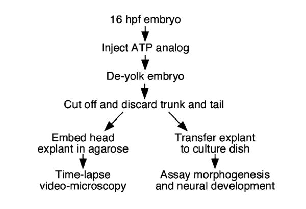
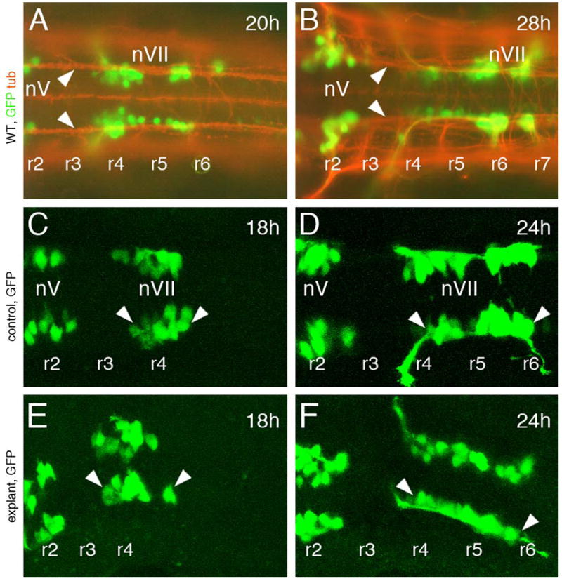
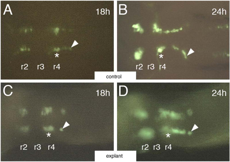
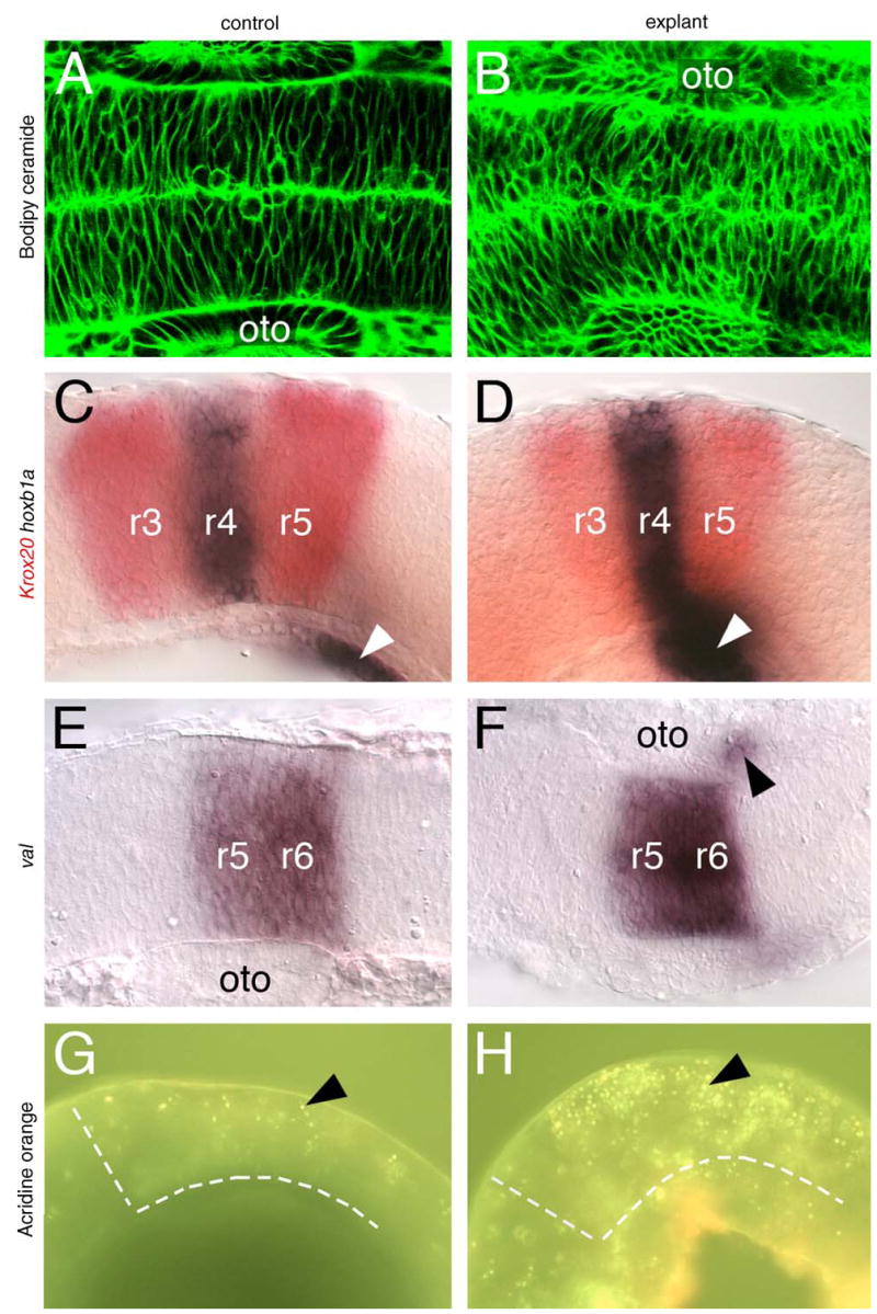
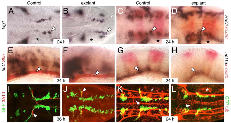
The zebrafish embryo is an excellent system for studying dynamic processes such as cell migration during vertebrate development. Dynamic analysis of neuronal migration in the zebrafish hindbrain has been hampered by morphogenetic movements in vivo, and by the impermeability of embryos. We have applied a recently reported technique of embryo explant culture to the analysis of neuronal development and migration in the zebrafish hindbrain. We show that hindbrain explants prepared at the somitogenesis stage undergo normal morphogenesis for at least 14 h in culture. Importantly, several aspects of hindbrain development such as patterning, neurogenesis, axon guidance, and neuronal migration are largely unaffected, inspite of increased cell death in explanted tissue. These results suggest that hindbrain explant culture can be employed effectively in zebrafish to analyze neuronal migration and other dynamic processes using pharmacological and imaging techniques.


Figures

Fig. 1
Fig. 1
Generation and analysis of hindbrain explants. Embryos were injected with the ATP analog (AMP-PNP) in the yolk cell. Following yolk removal, the embryo was decapitated, and the head fragment containing the hindbrain was either embedded in agarose for imaging and video microscopy or transferred to culture medium for assaying morphogenesis and neural development at subsequent stages. (see Section 2 for details).
Fig. 2
Fig. 2
Facial branchiomotor neurons (FBMNs) develop and migrate normally in hindbrain explants. All panels show dorsal views of the hindbrain, with anterior to the left. Rhombomere locations were assigned on the basis of their positions relative to the otic vesicle (not shown). A and B are composite images of GFP-expressing cells (green) in embryos labeled with an antibody against acetylated tubulin (orange). (A and B) In intact 20 and 28 hpf wild-type embryos, FBMNs (nVII motor neurons) are associated with axons of the ventral longitudinal fascicle (arrowheads) along the entire migratory pathway from rhombomere 4 (r4) to r5, r6 and r7. (C and E) In an intact (control) 18 hpf embryo embedded in agarose (C) and in an 18 hpf hindbrain explant, the FBMNs (nVII, arrowheads) are found in r4 and have just initiated caudal migration. (D and F) In a control 24 hpf embryo (D) and a 24 hpf hindbrain explant (F), several nVII motor neurons have migrated out of r4 into r5 and r6.
Fig. 3
Fig. 3
Video microscopy of migrating FBMNs in hindbrain explants. All panels show dorsal views of the hindbrain with anterior to the left. (A and B) In an intact 18 hpf control embryo (A), a GFP-expressing motor neuron (arrowhead) is close to its origin within r4 (asterisk). By 24 hpf (B), the same cell (arrowhead) has migrated more caudally into r6. (C and D) In an 18 hpf hindbrain explant (C), an isolated GFP-expressing motor neuron (arrowhead) is found in r5. By 24 hpf (D), the same cell (arrowhead) has migrated caudally into r6.
Fig. 4
Fig. 4
Morphogenesis and patterning is mostly normal in hindbrain explants. A, B, E, F show dorsal views, and C, D, G, H show lateral views of the hindbrain, with anterior to the left. (A and B) Bodipy-ceramide (green) labels the interstitial spaces between neuroepithelial cells, and shows that the cell shapes and their organization are comparable between an intact control embryo (A) and a hindbrain explant (B). (C–F) In control embryos, krox20 (red), hoxb1a (C), and val (E) are expressed in characteristic patterns in r3 and r5 (krox20), r4 (hoxb1a), and r5 and r6 (val), respectively. In hindbrain explants (D and F), these rhombomere-specific genes are expressed normally. Neural crest cells expressing val (arrowhead in F) migrate normally out of r6 in explants, and putative neural crest cells expressing hoxb1a (arrowheads in C and D) develop normally in explants. The apparently stronger and more anterior expression of hoxb1a in the explant (D) results from the curling and compression of the explanted tissue following deyolking (see panel H). (G and H) Acridine orange labels a few dying cells (arrowhead) in an intact control embryo (G). In a hindbrain explant (H), the number of dying cells (arrowhead) is significantly higher. The broken white lines in G and H delineate the anterior and ventral margins of the hindbrain. oto: Otocyst.
Fig. 5
Fig. 5
Neurogenesis and axonal patterning occur normally in hindbrain explants. A–D, and I–L show dorsal views, and E–H show lateral views of the hindbrain with anterior to the left. Asterisks in A–D, K and L mark the otocyst. (A and B) FBMNs (arrowhead) differentiate and migrate normally at 24 hpf in an intact control embryo (A) and a hindbrain explant (B). (C and D) Post-mitotic huC-expressing neurons (arrowhead) are distributed at the lateral margin of rhombomeres 3, 4, and 5 in a similar fashion in an intact control embryo (C) and a hindbrain explant (D). Krox20 expression (red) is evident in r3 and r5. (E and F) Floor plate cells (arrowhead) express shh in a similar manner in an intact control embryo (E) and a hindbrain explant (F). The number of huC-expressing cells is slightly increased in the explant. (G and H) Netrin1a, an axon guidance gene induced by Shh signaling, is expressed (arrowhead) throughout the ventral neural tube in an intact control embryo (G). Net1a expression is intact but slightly reduced in a hindbrain explant (H). (I and J) The Mauthner reticulospinal neurons (red, arrowhead) and their decussating axons exhibit similar morphologies at 36 hpf in an intact control embryo (I) and a hindbrain explant (J). (K and L) An antibody against acetylated tubulin labels longitudinal axon fasicles (red) and individual reticulospinal axons (arrowhead) in a similar fashion in an intact control embryo, and a hindbrain explant. Longitudinal axon fasicles on one side appear fused in the explant due to tilting of the tissue. GFP-expressing FBMNs migrate normally in explants (J and L) in a similar fashion to intact embryos (I and K).

Similar articles

Cited by

References

    1. Bingham S, Nasevicius A, Ekker SC, Chandrasekhar A. Sonic hedgehog and tiggy-winkle hedgehog cooperatively induce zebrafish branchiomotor neurons. Genesis. 2001;30:170–4. - PMC - PubMed
    1. Bingham S, Higashijima S, Okamoto H, Chandrasekhar A. The Zebrafish trilobite gene is essential for tangential migration of branchiomotor neurons. Dev Biol. 2002;242:149–60. - PMC - PubMed
    1. Bingham S, Chaudhari S, Vanderlaan G, Itoh M, Chitnis A, Chandrasekhar A. Neurogenic phenotype of mind bomb mutants leads to severe patterning defects in the zebrafish hindbrain. Dev Dyn. 2003;228:451–63. - PMC - PubMed
    1. Brand M, Heisenberg CP, Warga RM, Pelegri F, Karlstrom RO, Beuchle D, et al. Mutations affecting development of the midline and general body shape during zebrafish embryogenesis. Development. 1996;123:129–42. - PubMed
    1. Chandrasekhar A, Moens CB, Warren JT, Jr, Kimmel CB, Kuwada JY. Development of branchiomotor neurons in zebrafish. Development. 1997;124:2633–44. - PubMed

Publication types

MeSH terms

LinkOut - more resources