. 2005 Jun;2(6):e161.
doi: 10.1371/journal.pmed.0020161. Epub 2005 Jun 28.

Derivation of multipotent mesenchymal precursors from human embryonic stem cells

Affiliations

Derivation of multipotent mesenchymal precursors from human embryonic stem cells

Tiziano Barberi et al. PLoS Med. 2005 Jun.

Abstract

Background: Human embryonic stem cells provide access to the earliest stages of human development and may serve as a source of specialized cells for regenerative medicine. Thus, it becomes crucial to develop protocols for the directed differentiation of embryonic stem cells into tissue-restricted precursors.

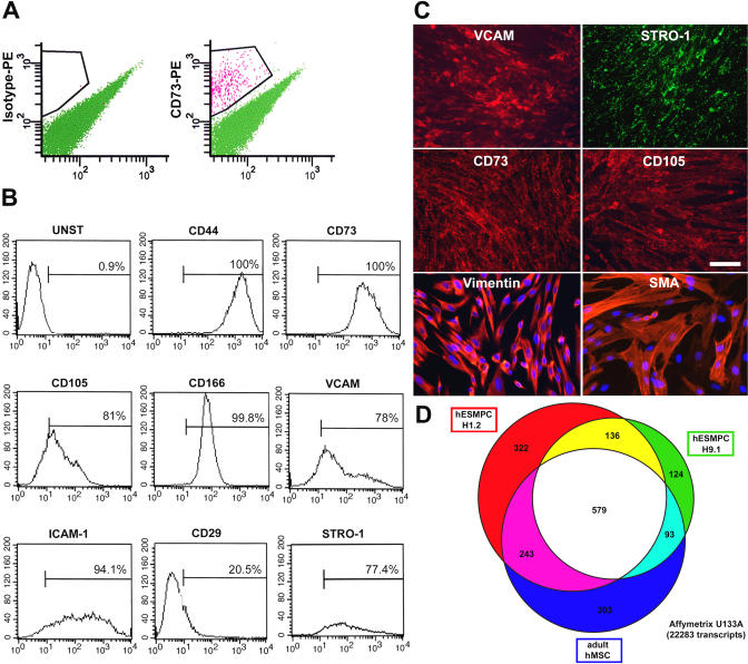
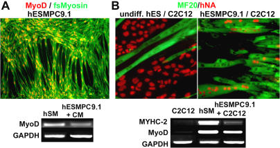
Methods and findings: Here, we present culture conditions for the derivation of unlimited numbers of pure mesenchymal precursors from human embryonic stem cells and demonstrate multilineage differentiation into fat, cartilage, bone, and skeletal muscle cells.

Conclusion: Our findings will help to elucidate the mechanism of mesoderm specification during embryonic stem cell differentiation and provide a platform to efficiently generate specialized human mesenchymal cell types for future clinical applications.


Conflict of interest statement

Competing Interests: The authors have declared that no competing interests exist.

Figures

Figure 1
Figure 1. Isolation and Characterization of hESMPCs
(A) FACS (MoFlo, Cytomation) for the isolation of CD73+ precursors (right) and isotype control (left). (B) Flow cytometry analysis of the CD73+ hESMPC population for various markers characteristic of MSCs, including CD44, CD73, CD105, CD166, VCAM, ICAM-1, CD29, and STRO-1. (C) Immunocytochemistry of hESMPCs for MSC markers (VCAM, STRO-1, CD73, and CD105). The cells also express vimentin and alpha smooth muscle actin. Scale bar = 50 μm. (D) Venn diagram presenting the overlap among transcripts selectively expressed in hESMPC-H1.2, hESMPC-H9.1, and primary adult human MSCs.
Figure 2
Figure 2. Selective Differentiation of hESMPCs into Various Mesenchymal Derivatives
(A) Adipocytic differentiation in the presence of dexamethasone, insulin, and isobutylxanthine. Adipocytic characterization by Oil Red O staining and RT-PCR analysis for PPARγ. (B) Chondrocytic differentiation in the presence of TGF-β3 and ascorbic acid. Chondrocytic characterization by Alcian Blue staining and RT-PCR for aggrecan and collagen II. (C) Osteogenic differentiation in the presence of β-glycerolphosphate, dexamethasone, and ascorbic acid. Osteocytic characterization by von Kossa staining and RT-PCR for bone-specific alkaline phosphatase (ALP) and bone sialoprotein (BSP). (D) Phase-contrast image of hESMPCs and RT-PCR for the ES cell markers Nanog and Oct-4 in hESMPC-H1.1 and -H9.1 compared with undifferentiated H1 hESCs. Scale bar = 50 μm for all panels.
Figure 3
Figure 3. Myogenic Differentiation of hESMPCs
(A) Immunocytochemistry for MyoD (red) and fast-switch myosin (green). RT-PCR for MyoD in human skeletal muscle as a positive control (hSM), and in hESMPC-H9.1 cells differentiated for 10 d in the presence of C2C12-conditioned medium (hESMPC). (B) Myotube formation induced at high cell densities in the presence of C2C12 cells. Myotube characterization by immunocytochemistry for MF20 against sarcomeric myosin (green) and human nuclear antigen (hNA, red). Left panel: Control undifferentiated hESCs (H9) do not fuse with C2C12. Right panel: Under identical culture conditions, hESMPCs (line 9.1) efficiently fuse with C2C12 cells, forming myotubes containing human nuclei. RT-PCR for human specific muscle transcripts myosin heavy chain IIa (MYHC-2) and MyoD in C2C12 cells, in human skeletal muscle as positive control (huSM), and in hESMPC-H9.1 cells cocultured with C2C12 cells.

References

    1. Perrier AL, Tabar V, Barberi T, Rubio ME, Bruses J, et al. Derivation of midbrain dopamine neurons from human embryonic stem cells. Proc Natl Acad Sci U S A. 2004;101:12543–12548. - PMC - PubMed
    1. Reubinoff BE, Itsykson P, Turetsky T, Pera MF, Reinhartz E, et al. Neural progenitors from human embryonic stem cells. Nat Biotechnol. 2001;19:1134–1140. - PubMed
    1. Zhang SC, Wernig M, Duncan ID, Brustle O, Thomson JA. In vitro differentiation of transplantable neural precursors from human embryonic stem cells. Nat Biotechnol. 2001;19:1129–1133. - PubMed
    1. Sottile V, Thomson A, McWhir J. In vitro osteogenic differentiation of human ES cells. Cloning Stem Cells. 2003;5:149–155. - PubMed
    1. Pittenger MF, Mackay AM, Beck SC, Jaiswal RK, Douglas R, et al. Multilineage potential of adult human mesenchymal stem cells. Science. 1999;284:143–147. - PubMed

Publication types

MeSH terms