. 2005 Oct 15;391(Pt 2):291-300.
doi: 10.1042/BJ20050468.

The selective recruitment of mRNA to the ER and an increase in initiation are important for glucose-stimulated proinsulin synthesis in pancreatic beta-cells

Affiliations

The selective recruitment of mRNA to the ER and an increase in initiation are important for glucose-stimulated proinsulin synthesis in pancreatic beta-cells

Isabel C Greenman et al. Biochem J. .

Abstract

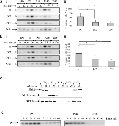
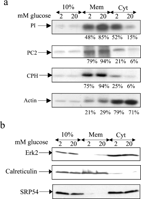
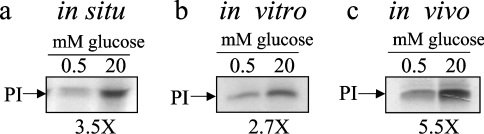
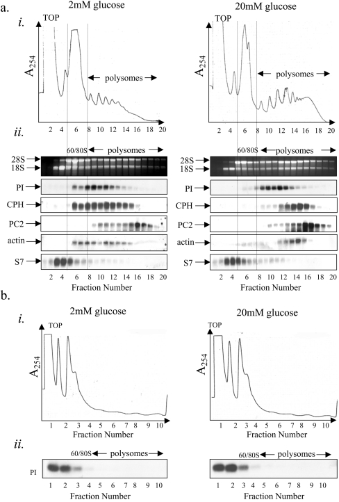
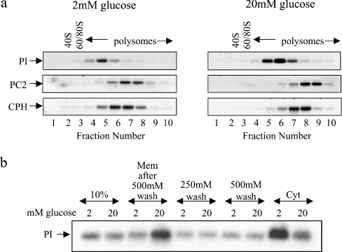
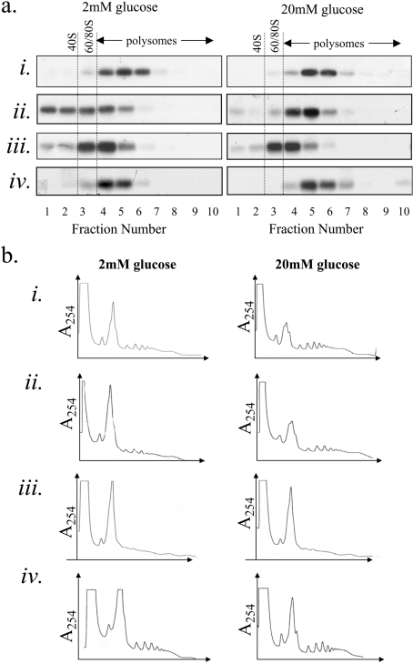
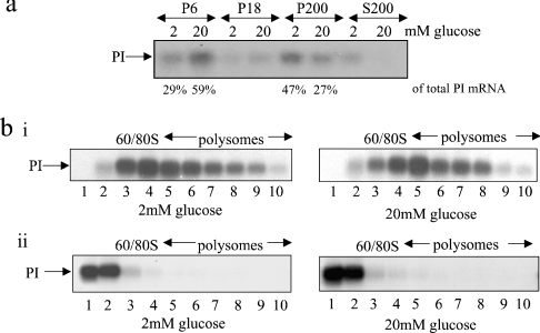
Glucose acutely stimulates proinsulin synthesis in pancreatic beta-cells through a poorly understood post-transcriptional mechanism. In the present study, we demonstrate in pancreatic beta-cells that glucose stimulates the recruitment of ribosome-associated proinsulin mRNA, located in the cytoplasm, to the ER (endoplasmic reticulum), the site of proinsulin synthesis, and that this plays an important role in glucose-stimulated proinsulin synthesis. Interestingly, glucose has greater stimulatory effect on the recruitment of proinsulin mRNA to the ER compared with other mRNAs encoding secretory proteins. This, as far as we are aware, is the first example whereby mRNAs encoding secretory proteins are selectively recruited to the ER and provides a novel regulatory mechanism for secretory protein synthesis. Contrary to previous reports, and importantly in understanding the mechanism by which glucose stimulates proinsulin synthesis, we demonstrate that there is no large pool of 'free' proinsulin mRNA in the cytoplasm and that glucose does not increase the rate of de novo initiation on the proinsulin mRNA. However, we show that glucose does stimulate the rate of ribosome recruitment on to ribosome-associated proinsulin mRNA. In conclusion, our results provide evidence that the selective recruitment of proinsulin mRNA to the ER, together with increases in the rate of initiation are important mediators of glucose-stimulated proinsulin synthesis in pancreatic beta-cells.


Figures

Figure 1
Figure 1. Glucose stimulates the recruitment of mRNAs encoding secretory membrane proteins to the ER: subcellular fractionation by consecutive centrifugation
MIN6 cells were preincubated in KRB containing 0.5 or 2 mM glucose for 1 h followed by incubation for a further period of 1 h at the same glucose concentration or 20 mM glucose. Cells were fractionated by consecutive centrifugation resulting in a membrane fraction (P6), a residual membrane fraction (P18), a polysome fraction (P200) and a supernatant fraction containing free mRNAs (S200); 10% refers to 10% of total RNA/protein. (ai, bi) RNA isolated from each fraction was run on a 1% agarose formaldehyde gel, transferred on to a nylon membrane and probed for proinsulin (PI), PC2, CPH and actin mRNAs. (aii, bii) The percentage increase in mRNA levels at the ER in response to high glucose was determined by quantification of bands from Northern-blot analysis using ImageJ. The error bars indicate the S.E.M. (n=7). Significant differences are indicated by *P<0.05 (two sample t test). (c) Protein samples from each fraction (8% of the P6/P18/P200 fraction and 1% of the S200 fraction) were run on SDS/PAGE (12.5% polyacrylamide) and subjected to immunoblotting with anti-ERK2, calreticulin and SRP54 antibodies. Detection was by enhanced chemiluminescence. (d) Time course of the recruitment of proinsulin mRNA to the ER. MIN6 cells were incubated in KRB containing 0.5 mM glucose followed by incubation for the times indicated in KRB containing 20 mM glucose. Results presented are representative of three separate experiments.
Figure 2
Figure 2. Glucose stimulates the recruitment of mRNAs encoding secretory membrane proteins to the ER: subcellular fractionation using the digitonin method
MIN6 cells were preincubated in KRB containing 2 mM glucose for 1 h followed by incubation in KRB containing 2 or 20 mM glucose for a further period of 1 h. (a) Cells were permeabilized with digitonin and pelleted resulting in a membrane fraction (Mem) and a cytosolic fraction (Cyt). The 10% refers to 10% of total RNA/protein. RNA was isolated from each fraction and run on a 1% agarose formaldehyde gel, transferred on to a nylon membrane and probed for proinsulin (PI), PC2, CPH and actin mRNAs. The intensities of the bands were quantified using ImageJ and the mRNA levels were expressed as a percentage of total mRNA. (b) Protein samples from each fraction were run on SDS/PAGE (20% of membrane fraction and 20% of cytosolic fraction) and subjected to immunoblotting with anti-ERK2, calreticulin and SRP54 antibodies. Detection was by enhanced chemiluminescence. The results presented are representative of three separate experiments.
Figure 3
Figure 3. The recruitment of proinsulin mRNA on to the membranes at high glucose plays a significant role in glucose-stimulated proinsulin synthesis
MIN6 cells were preincubated in KRB containing 0.5 mM glucose for 1 h followed by incubation in KRB containing 0.5 or 20 mM glucose for a further period of 1 h. Membranes were isolated by consecutive centrifugation as described in Figure 1. (a) Resuspended membranes were translated in situ in the presence of [35S]methionine. After translation, the membranes were pelleted at 13000 g for 10 min. Proinsulin (PI) was resolved by SDS/PAGE. (b) Poly(A)+ selected mRNA was isolated from the membranes and translated in vitro in the presence of [35S]methionine. Proinsulin (PI) was immunoprecipitated and resolved by SDS/PAGE. (c) MIN6 cells were preincubated in KRB containing 0.5 mM glucose for 1 h followed by incubation in KRB containing 0.5 or 20 mM glucose in the presence of [35S]methionine for a further period of 1 h. The cells were lysed and proinsulin (PI) resolved by SDS/PAGE. In all cases, proteins were visualized by autoradiography. In (a, c) the results are representative of three separate experiments and in (b) the results are representative of two separate experiments.
Figure 4
Figure 4. Glucose stimulates an increase in ribosome recruitment on to ribosome-associated proinsulin mRNA
(a) MIN6 cells were preincubated in KRB containing 2 mM glucose for 1 h followed by incubation in KRB containing 2 or 20 mM glucose for a further period of 1 h. Cells were lysed and polysome analysis was carried out using 7–47% sucrose gradients. The gradients were fractionated from left (fraction 1) to right (fraction 20). (ai) Absorbance of the gradients was measured continually at 254 nm to give polysome profiles. (aii) RNA was isolated from each fraction and run on a 1% agarose formaldehyde gel. RNA was transferred on to a nylon membrane and probed for proinsulin (PI), CPH, PC2, actin and S7 mRNAs. The results presented are representative of three separate experiments. (b) Cells were treated and fractionated as described in (a) except that 15 mM EDTA was included in the lysis buffer and the sucrose gradients to dissociate the ribosomes.
Figure 5
Figure 5. Glucose stimulates the recruitment of ribosomes on to membrane-bound proinsulin mRNA
MIN6 cells were preincubated in KRB containing 2 mM glucose for 1 h followed by incubation in KRB containing 2 or 20 mM glucose for a further period of 1 h. (a) Cells were then lysed and fractionated by consecutive centrifugation. The membrane fractions were layered on to 20–50% sucrose gradients, centrifuged and fractionated from left (fraction 1) to right (fraction 10). RNA was isolated from each fraction and run on a 1% agarose formaldehyde gel. RNA was then transferred on to a nylon membrane and probed for proinsulin (PI), PC2 and CPH mRNAs. (b) Cells were then lysed and fractionated by the digitonin cell permeabilization method (using modified polysome buffer containing 130 mM KCl), resulting in a membrane fraction (Mem) and a cytosolic fraction (Cyt). The membrane fraction was consecutively washed with polysome buffer containing 250 mM KCl (250 mM wash) and 500 mM KCl (500 mM wash). RNA isolated from each fraction and washed was run on a 1% agarose formaldehyde gel, transferred on to a nylon membrane and probed for proinsulin (PI) mRNA; 10% refers to 10% of total RNA.
Figure 6
Figure 6. Evidence for increased ribosome recycling/recruitment on to proinsulin mRNA at high glucose
MIN6 cells were preincubated in KRB containing 2 mM glucose for 1 h followed by incubation in KRB containing 2 or 20 mM glucose for a further period of 1 h. During the final 10 min of incubation, the following were added: (ai, bi) 0.1 mg/ml cycloheximide, (aii, bii) no treatment, (aiii, biii) 0.1 μg/ml pactamycin and (aiv, biv) 0.1 mg/ml cycloheximide and 0.1 μg/ml pactamycin. Cells were lysed and polysome analysis was carried out on 20–50% sucrose gradients. Gradients were fractionated from left (fraction 1) to right (fraction 10). (a) Northern-blot analysis: RNA was isolated from each fraction and run on a 1% agarose formaldehyde gel. RNA was then transferred on to a nylon membrane and probed for proinsulin (PI) mRNA. (b) Polysome profiles.
Figure 7
Figure 7. Glucose-stimulated proinsulin synthesis in islets of Langerhans
Rat islets were preincubated in KRB containing 2 mM glucose for 2 h followed by incubation in KRB containing 2 or 20 mM glucose for a further period of 1 h. (a) Cells were fractionated by consecutive centrifugation as described in Figure 1. RNA was isolated from each fraction and run on a 1% agarose formaldehyde gel, then transferred on to a nylon membrane and probed for proinsulin (PI) mRNA; (bi) Cells were lysed and the lysates were layered on to 20–50% sucrose gradients, centrifuged and fractionated from left (fraction 1) to right (fraction 10). RNA was isolated from each fraction and run on a 1% agarose formaldehyde gel. RNA was then transferred on to a nylon membrane and probed for proinsulin (PI) mRNA. (bii) Cells were treated and fractionated as described in (bi) except that 15 mM EDTA was included in the lysis buffer and the sucrose gradients to dissociate the ribosomes but leave mRNPs intact.

References

    1. Campbell I. L., Hellquist L. N., Taylor K. W. Insulin biosynthesis and its regulation. Clin. Sci. 1982;62:449–455. - PubMed
    1. Itoh N., Sei T., Nose K., Okamoto H. Glucose stimulation of the proinsulin synthesis in isolated pancreatic islets without increasing amount of proinsulin mRNA. FEBS Lett. 1978;93:343–347. - PubMed
    1. Itoh N., Okamoto H. Translational control of proinsulin synthesis by glucose. Nature (London) 1980;283:100–102. - PubMed
    1. Permutt M. A., Kipnis D. M. Insulin biosynthesis. I. On the mechanism of glucose stimulation. J. Biol. Chem. 1972;247:1194–1199. - PubMed
    1. Jahr H., Schroder D., Ziegler B., Ziegler M., Zuhlke H. Transcriptional and translational control of glucose-stimulated (pro)insulin biosynthesis. Eur. J. Biochem. 1980;110:499–505. - PubMed

Publication types

MeSH terms