. 2005 Jun 22;6(1):61.
doi: 10.1186/1465-9921-6-61.

Alveolar macrophages regulate neutrophil recruitment in endotoxin-induced lung injury

Affiliations

Alveolar macrophages regulate neutrophil recruitment in endotoxin-induced lung injury

Beatrice Beck-Schimmer et al. Respir Res. .

Abstract

Background: Alveolar macrophages play an important role during the development of acute inflammatory lung injury. In the present study, in vivo alveolar macrophage depletion was performed by intratracheal application of dichloromethylene diphosphonate-liposomes in order to study the role of these effector cells in the early endotoxin-induced lung injury.

Methods: Lipopolysaccharide was applied intratracheally and the inflammatory reaction was assessed 4 hours later. Neutrophil accumulation and expression of inflammatory mediators were determined. To further analyze in vivo observations, in vitro experiments with alveolar epithelial cells and alveolar macrophages were performed.

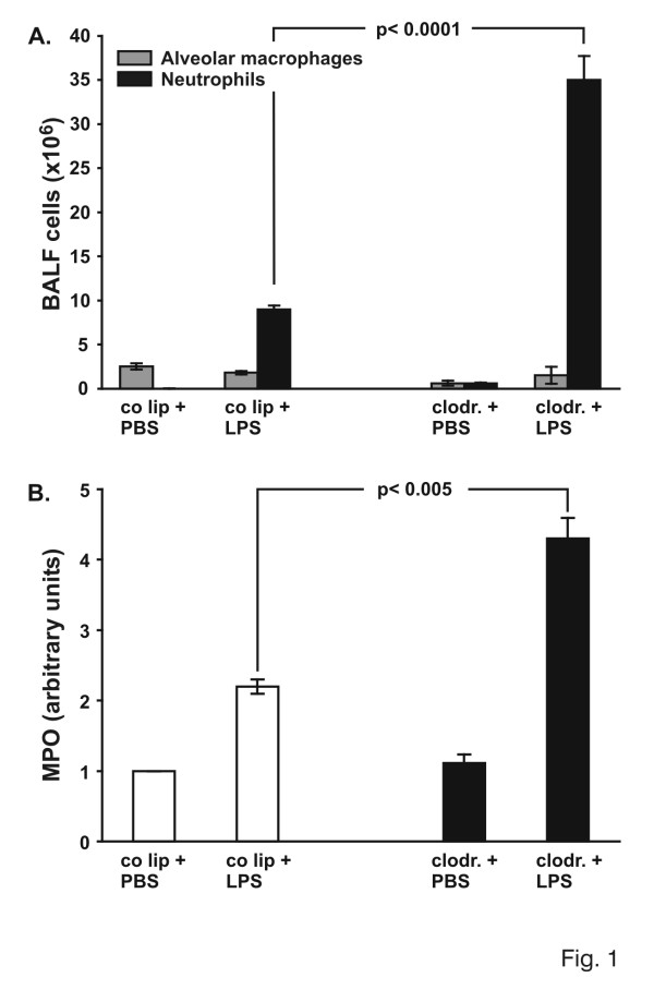
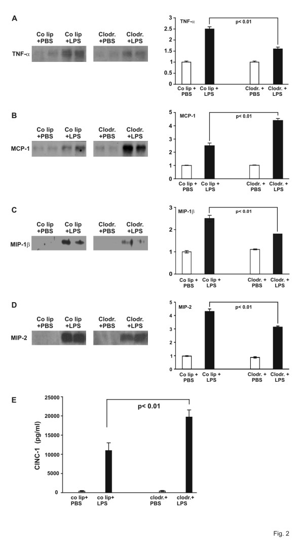
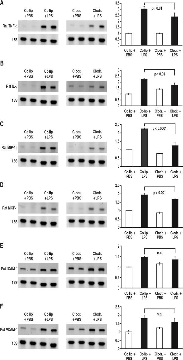
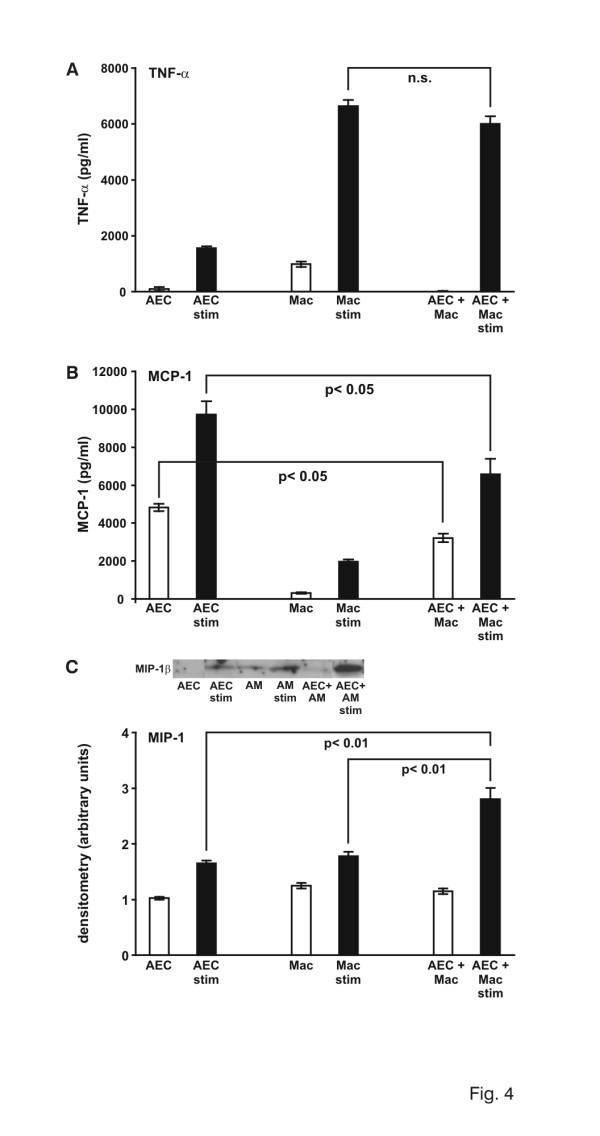
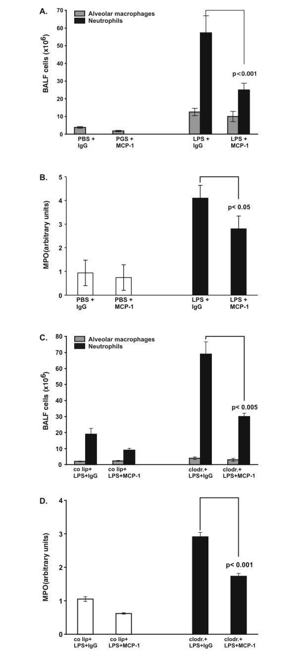
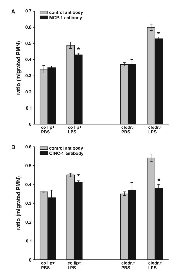
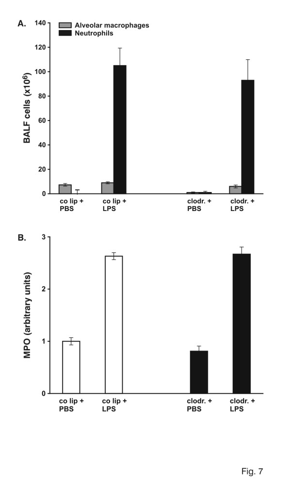
Results: A 320% increase of polymorphonuclear leukocytes in bronchoalveolar lavage fluid was observed in macrophage-depleted compared to macrophage-competent lipopolysaccharide-animals. This neutrophil recruitment was also confirmed in the interstitial space. Monocyte chemoattractant protein-1 concentration in bronchoalveolar lavage fluid was significantly increased in the absence of alveolar macrophages. This phenomenon was underlined by in vitro experiments with alveolar epithelial cells and alveolar macrophages. Neutralizing monocyte chemoattractant protein-1 in the airways diminished neutrophil accumulation.

Conclusion: These data suggest that alveolar macrophages play an important role in early endotoxin-induced lung injury. They prevent neutrophil influx by controlling monocyte chemoattractant protein-1 production through alveolar epithelial cells. Alveolar macrophages might therefore possess robust anti-inflammatory effects.


Figures

Figure 1
Figure 1
A: Total cell count in bronchoalveolar lavage fluid (BALF). Animals were pretreated with control liposomes (co lip) or clodronate-liposomes (clodronate). 72 hours later 150 μg LPS was instilled intratracheally and lungs were evaluated 4 hours later. Cells were analyzed using cytospin and Diff-Quick staining. Values are mean ± SEM from 5 animals per group. B. Determination of myeloperoxidase (MPO) activity as a measure of parenchymal neutrophil content. Animals were pretreated with control liposomes (co lip) or clodronate-liposomes (clodronate). 72 hours later 150 μg LPS was instilled intratracheally and lungs were evaluated 4 hours later. The right lung of each animal was removed and homogenized in sample buffer. Optical density was measured at 420 nm over 360s. The value for control liposomes and PBS was defined as 1 and all other values were adapted. Values are mean ± SEM from 5 animals per group.
Figure 2
Figure 2
A. TNF-α and protein determination in bronchoalveolar lavage fluid of AM-competent (control liposome = co lip) and AM-depleted (clodronate) animals, exposed to PBS or LPS for 4 hours. Proteins were electorphoresed on a SDS-polyacrylamide gel and transblotted to a nitrocellulose membrane. Densitometry was performed. One value for control liposomes and PBS was defined as 1 and all other values were adapted. Values are mean ± SEM from 5 animals (only 2 are shown). B. MCP-1 protein determination in bronchoalveolar lavage fluid of AM-competent and -depleted animals, exposed to PBS or LPS for 4 hours. Proteins were electorphoresed on a SDS-polyacrylamide gel and transblotted to a nitrocellulose membrane. Densitometry was performed. One value for control liposomes and PBS was defined as 1 and all other values were adapted. Values are mean ± SEM from 5 animals (only 2 are shown). C. MIP-1β protein determination in bronchoalveolar lavage fluid of AM-competent and -depleted animals, exposed to PBS or LPS for 4 hours. Proteins were electorphoresed on a SDS-polyacrylamide gel and transblotted to a nitrocellulose membrane. Densitometry was performed. One value for control liposomes and PBS was defined as 1 and all other values were adapted. Values are mean ± SEM from 5 animals (only 2 are shown). D. MIP-2 protein determination in bronchoalveolar lavage fluid of AM-competent and -depleted animals, exposed to PBS or LPS for 4 hours. Proteins were electorphoresed on a SDS-polyacrylamide gel and transblotted to a nitrocellulose membrane. Densitometry was performed. One value for control liposomes and PBS was defined as 1 and all other values were adapted. Values are mean ± SEM from 5 animals (only 2 are shown). E. CINC concentration in bronchoalveolar lavage fluid (BALF) of AM-competent (control liposomes = co lip) and AM-depleted (clodronate) animals, exposed to PBS or LPS for 4 hours. CINC was examined using a standard ELISA. Values are mean ± SEM from 5 animals.
Figure 3
Figure 3
Changes in expression of TNF-α mRNA (A), IL-1β mRNA (B), MIP-1β mRNA (C), MCP-1 mRNA (D), ICAM-1 mRNA (E) and VCAM-1 mRNA (F) of AM-competent (control liposomes = co lip) and -depleted (clodrondate) animals after 4 hours of PBS/LPS stimulation. Whole lung RNA was extracted and RT-PCR performed according to the primers and annealing thermocycle conditions shown in Tab. 1. Equal loading was shown with 18S bands. Results were analyzed by densitometry. One value for control liposomes and PBS was defined as 1 and all other values were adapted. Values are mean ± SEM from 5 animals.
Figure 4
Figure 4
A. TNF-α protein determination in supernatants of alveolar epithelial cells (AEC), alveolar macrophages (AM) and co-culture of AEC/AM upon LPS stimulation (relation AEC:AM = 2:1). TNF-α was examined using a standard ELISA. White bars represent control cells, black bars LPS-stimulated cells. Values are mean ± SEM from 5 different assays. B. MCP-1 protein determination in supernatants of alveolar epithelial cells (AEC), alveolar macrophages (AM) and co-culture of AEC/AM upon LPS stimulation (relation AEC:AM = 2:1). MCP-1 was examined using a standard ELISA. White bars represent control cells, black bars LPS-stimulated cells. Values are mean ± SEM from 5 different assays. C. MIP-1β protein determination in supernatants of alveolar epithelial cells (AEC), alveolar macrophages (AM) and co-culture of AEC/AM upon LPS stimulation (relation AEC:AM = 2:1). MIP-1β was examined using Western blot analysis. Densitometry was performed. One value for control liposomes and PBS was defined as 1 and all other values were adapted. White bars represent control cells, black bars LPS-stimulated cells. Values are mean ± SEM from 5 different assays.
Figure 5
Figure 5
A. Total cell count in bronchoalveolar lavage fluid (BALF). Rats were challenged with intratracheal instillation of LPS or PBS, followed by intratracheal application of MCP-1 antibody or a control antibody (IgG). Lungs were evaluated 4 hours later. Cells were analyzed using cytospin and Diff-Quick staining. Values are mean ± SEM from 5 animals. B. Determination of myeloperoxidase (MPO) activity as a measure of parenchymal PMN content. Rats were challenged with intratracheal instillation of LPS or PBS, followed by intratracheal application of MCP-1 antibody or a control antibody (IgG). Lungs were evaluated 4 hours later. The right lung of each animal was removed and homogenized in sample buffer. Optical density was measured at 420 nm over 360s. One value PBS+IgG was defined as 1 and all other values were adapted. Values are mean ± SEM from 5 animals. C. Total cell count in bronchoalveolar lavage fluid (BALF). AM-competent (control liposomes = co lip) and -depleted (clodrondate) animals were challenged with intratracheal instillation of LPS, followed by intratracheal application of MCP-1 antibody or a control antibody (IgG). Lungs were evaluated 4 hours later. Cells were analyzed using cytospin and Diff-Quick staining. Values are mean ± SEM from 5 animals. D. Determination of myeloperoxidase (MPO) activity as a measure of parenchymal PMN content. AM-competent (control liposomes = co lip) and -depleted (clodrondate) animals were challenged with intratracheal instillation of LPS, followed by intratracheal application of MCP-1 antibody or a control antibody (IgG). Lungs were evaluated 4 hours later. The right lung of each animal was removed and homogenized in sample buffer. Optical density was measured at 420 nm over 360s. One value LPS+IgG was defined as 1 and all other values were adapted. Values are mean ± SEM from 5 animals.
Figure 6
Figure 6
A. Chemotaxis assays performed with BALF of control liposome-PBS/LPS and clodronate-PBS/LPS animals. 1 × 10⁵ polymorphonuclear cells (PMN) were labelled with calcein and given to the upper chamber, while the lower one was loaded with 150 μl of BALF. 10 μg/ml neutralizing anti-rat MCP-1 antibody or 10 μg/ml control IgG were added to BALF. PBS/1% fetal bovine serum was taken to determine basal migration. Migrated cells were lysed and fluorescence was measured by using an excitation filter at 485 nm and an emission filter at 535 nm. Ratio between stimulated and basal migration was used as indication of PMN chemotaxis. * p < 0.05 B. Chemotaxis assays performed with BALF of control liposome-PBS/LPS and clodronate-PBS/LPS animals. 1 × 10⁵ polymorphonuclear cells (PMN) were labelled with calcein and given to the upper chamber, while the lower one was loaded with 150 μl of BALF. 10 μg/ml neutralizing anti-rat CINC-1 antibody or 10 μg/ml MOPC-21 were added to BALF. PBS/1% fetal bovine serum was taken to determine basal migration. Migrated cells were lysed and fluorescence was measured by using an excitation filter at 485 nm and an emission filter at 535 nm. Ratio between stimulated and basal migration was used as indication of PMN chemotaxis. * p < 0.05
Figure 7
Figure 7
A: Total cell count in bronchoalveolar lavage fluid (BALF). Animals were pretreated with control liposomes (co lip) or clodronate-liposomes (clodronate). 72 hours later 150 μg LPS was instilled intratracheally and lungs were evaluated 24 hours later. Cells were analyzed using cytospin and Diff-Quick staining. Values are mean ± SEM from 5 animals. B. Determination of myeloperoxidase (MPO) activity as a measure of parenchymal neutrophil content. Animals were pretreated with control liposomes (co lip) or clodronate-liposomes (clodronate). 72 hours later 150 μg LPS was instilled intratracheally and lungs were evaluated 24 hours later. The right lung of each animal was removed and homogenized in sample buffer. Optical density was measured at 420 nm over 360s. One value for control liposomes and PBS was defined as 1 and all other values were adapted. Values are mean ± SEM from 5 animals.

Similar articles

Cited by

References

    1. Beck-Schimmer B, Schimmer RC, Warner RL, Schmal H, Nordblom G, Flory CM, Lesch ME, Friedl HP, Schrier DJ, Ward PA. Expression of lung vascular and airway ICAM-1 after exposure to bacterial lipopolysaccharide. Am J Respir Cell Mol Biol. 1997;17:344–352. - PubMed
    1. Li XY, Donaldson K, MacNee W. Lipopolysaccharide-induced alveolar epithelial permeability: the role of nitric oxide. Am J Respir Crit Care Med. 1998;157:1027–1033. - PubMed
    1. Madjdpour C, Oertli B, Ziegler U, Bonvini JM, Pasch T, Beck-Schimmer B. Lipopolysaccharide induces functional ICAM-1 expression in rat alveolar epithelial cells in vitro. Am J Physiol Lung Cell Mol Physiol. 2000;278:L572–9. - PubMed
    1. Sibille Y, Reynolds HY. Macrophages and polymorphonuclear neutrophils in lung defense and injury. Am Rev Respir Dis. 1990;141:471–501. - PubMed
    1. Monton C, Torres A. Lung inflammatory response in pneumonia. Monaldi Arch Chest Dis. 1998;53:56–63. - PubMed

Publication types