. 2005 Jun 28;102(26):9300-5.
doi: 10.1073/pnas.0409873102. Epub 2005 Jun 21.

NF-kappaB activation and potentiation of proinflammatory responses by the Helicobacter pylori CagA protein

Affiliations

NF-kappaB activation and potentiation of proinflammatory responses by the Helicobacter pylori CagA protein

Sabine Brandt et al. Proc Natl Acad Sci U S A. .

Abstract

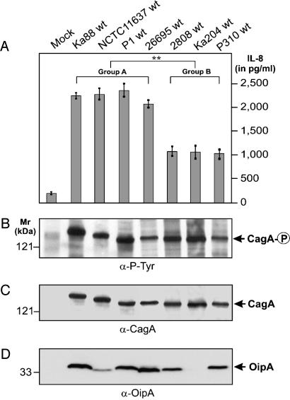
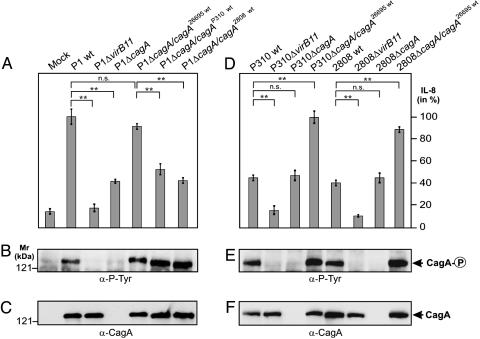
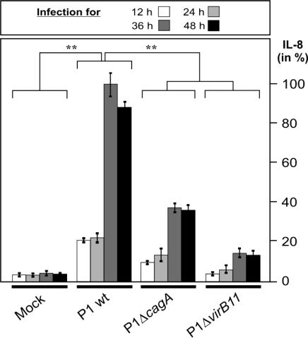
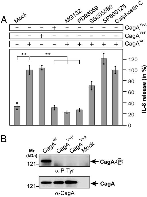
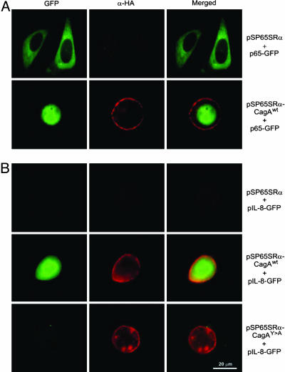
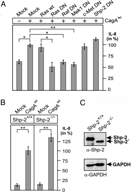
The Helicobacter pylori immunodominant protein, CagA, is associated with severe gastritis and carcinoma. Injection of CagA into gastric epithelial cells by type IV secretion leads to actin-cytoskeletal rearrangements and cell scattering. CagA has been reported to have no role in the induction of transcription factor NF-kappaB and IL-8, which are crucial determinants for chronic inflammation. Here, we provide several lines of evidence showing that CagA is able to induce IL-8 in a time- and strain-dependent manner. We also show that by exchanging specific cagA genes, high IL-8-inducing H. pylori strains could be converted into low inducing strains and vice versa. Our results suggest that IL-8 release induced by CagA occurs via a Ras-->Raf-->Mek-->Erk-->NF-kappaB signaling pathway in a Shp-2- and c-Met-independent manner. Thus, CagA is a multifunctional protein capable of effecting both actin remodeling and potentiation of chemokine release.


Figures

Fig. 1.
Fig. 1.
Identification of high and low IL-8-inducers among H. pylori strains that carry a functional cagPAI. (A) IL-8 release into the culture supernatant of AGS cells infected with various H. pylori strains was measured by standard ELISA. (B) Translocation and phosphorylation of CagA was analyzed by Western blotting with the phosphotyrosine-specific antibody PY-99. Stripping and reprobing of the blot with an α-CagA (C) or an α-OipA (D) antibody is shown. Arrows indicate the positions of the different proteins on the gel. Infection was for 9 h at a multiplicity of infection of 100. For the mock control, PBS was added to AGS. The data are mean values ± SD from at least three independent experiments.
Fig. 2.
Fig. 2.
CagA is involved in IL-8 induction. Genetic exchange of cagA genes reveals that low IL-8 inducers can be converted into high IL-8 inducers and vice versa. (A and D) ELISA reveals that exchange of cagA genes could alter the ability to induce IL-8 release. The cagA genes were exchanged between high and low IL-8-inducing H. pylori strains with the use of the shuttle plasmid pSB19. (B and E) CagA translocation and tyrosine phosphorylation in infected AGS cells was analyzed by Western blotting with antibody PY-99. (C and F) Stripping and reprobing of the blots with an α-CagA antibody. Arrows indictate the position of the different CagA protein species on the gel. Infection was for 9 h at a multiplicity of infection of 100. The data are mean values ± SD from at least three independent experiments.
Fig. 3.
Fig. 3.
CagA-mediated induction of IL-8 release is time-dependent and potentiates during the course of infection. ELISA reveals that the amount of IL-8 induced by WT H. pylori enhances in a cagPAI- and CagA-dependent manner during the course of infection at a multiplicity of infection of 50.
Fig. 4.
Fig. 4.
Induction of IL-8 by transfection of CagA. (A) WT and mutant CagA from the high-inducing H. pylori strain NCTC11637 were transiently expressed in AGS in the presence or absence of the proteasome inhibitor MG132 (30 μM), Erk inhibitor PD98059 (25 μM), MAP kinase p38 inhibitor SB203580 (25 μM), c-Jun N-terminal kinase inhibitor SP600125 (25 μM), and PKC inhibitor calphostin C (100 nM) as indicated. At 48 h after infection, IL-8 release into the culture supernatant was measured by standard ELISA. For the mock control, empty vector was transfected. The data are mean values ± SD from at least three independent experiments. (B) Expression and tyrosine phosphorylation of CagA were verified by Western blot analysis.
Fig. 5.
Fig. 5.
Immunofluorescence of AGS cells transfected with (A) NF-κB p65 subunit (p65-GFP, green) or (B) pIL-8-GFP (green) in the presence of HA-tagged CagA constructs expressed from vector pSP65SRα (α-HA-staining, red). Transient expression of CagAWT triggered both translocation of p65-GFP into the nucleus as well as induction of pIL-8-GFP, neither of which was seen with the empty pSP65SRα vector control. The CagAY→A mutant also did not induce pIL-8-GFP (B) or nuclear translocation of p65-GFP (data not shown).
Fig. 6.
Fig. 6.
Induction of IL-8 by CagA involves Ras, Raf, and Mek, but not Shp-2 or c-Met. (A) IL-8 release in AGS cells transfected with WT CagA in the presence of either WT or DN constructs. (B) IL-8 release in Shp-2-/- and Shp2+/+ cells transfected with CagAWT. As mock control, empty vector was transfected into the cells. The data are mean values ± SD from at least three independent experiments. (C) Expression of Shp-2 was verified by Western blot analysis with GAPDH as loading control.

Similar articles

Cited by

References

    1. Medzhitov, R. (2001) Nat. Rev. Immunol. 1, 135-145. - PubMed
    1. Akira, S. (2003) Curr. Opin. Immunol. 15, 5-11. - PubMed
    1. Beutler, B. (2004) Nature 430, 257-263. - PubMed
    1. Girardin, S. E., Sansonetti, P. J. & Philpott, D. J. (2002) Trends Microbiol. 10, 193-199. - PubMed
    1. Viala, J., Chaput, C., Boneca, I. G., Cardona, A., Girardin, S. E., Moran, A. P., Athman, R., Memet, S., Huerre, M. R., Coyle, A. J., et al. (2004) Nat. Immunol. 5, 1166-1174. - PubMed

Publication types

MeSH terms

Associated data