Comparative Study
. 2005 Oct;171(2):443-55.
doi: 10.1534/genetics.105.042101. Epub 2005 Jun 21.

A role for the Saccharomyces cerevisiae regulation of Ace2 and polarized morphogenesis signaling network in cell integrity

Affiliations
Comparative Study

A role for the Saccharomyces cerevisiae regulation of Ace2 and polarized morphogenesis signaling network in cell integrity

Cornelia Kurischko et al. Genetics. 2005 Oct.

Abstract

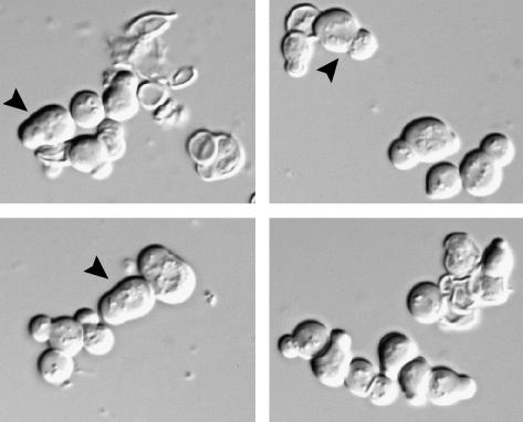
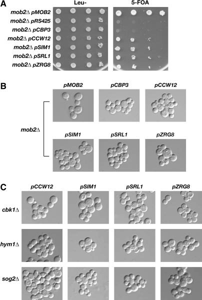
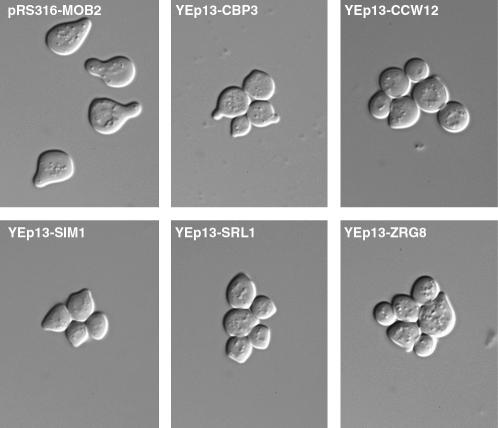
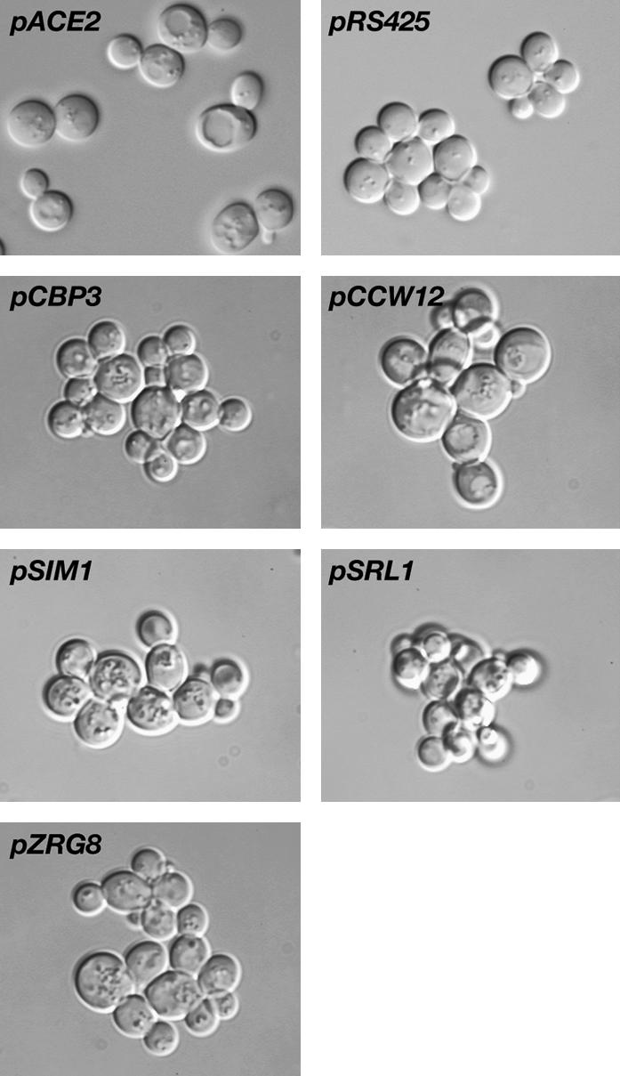
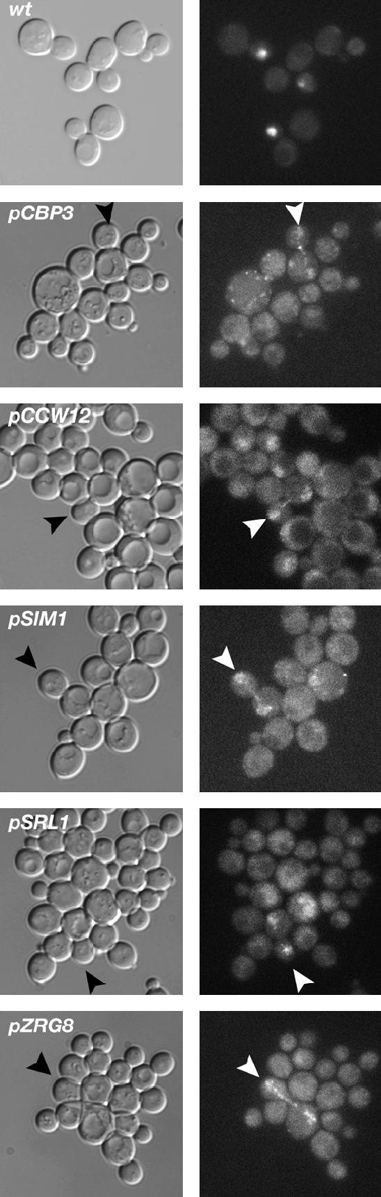
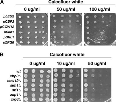
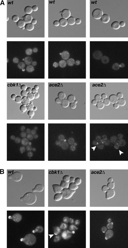
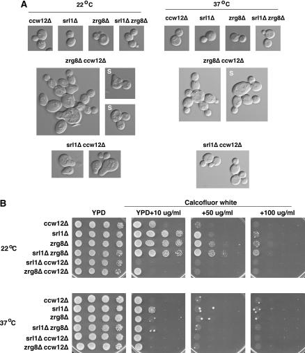
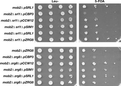
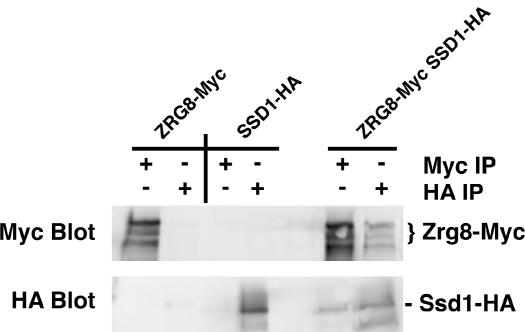
Saccharomyces cerevisiae RAM is a conserved signaling network that regulates maintenance of polarized growth and daughter-cell-specific transcription, the latter of which is critical for septum degradation. Consequently, cells defective in RAM function (designated ramDelta) are round in morphology, form feeble mating projections, and fail to separate following cytokinesis. It was recently demonstrated that RAM genes are essential in strains containing functional SSD1 (SSD1-v), which encodes a protein of unknown function that binds the RAM Cbk1p kinase. Here we investigated the essential function of RAM in SSD1-v strains and identified two functional groups of dosage suppressors for ramDelta lethality. We establish that all ramDelta mutants exhibit cell integrity defects and cell lysis. All dosage suppressors rescue the lysis but not the cell polarity or cell separation defects of ramDelta cells. One class of dosage suppressors is composed of genes encoding cell wall proteins, indicating that alterations in cell wall structure can rescue the cell lysis in ramDelta cells. Another class of ramDelta dosage suppressors is composed of ZRG8 and SRL1, which encode two unrelated proteins of unknown function. We establish that ZRG8 and SRL1 share similar genetic interactions and phenotypes. Significantly, Zrg8p coprecipitates with Ssd1p, localizes similarly to RAM proteins, and is dependent on RAM for localization. Collectively, these data indicate that RAM and Ssd1p function cooperatively to control cell integrity and suggest that Zrg8p and Srl1p function as nonessential inhibitors of Ssd1p.


Figures

Figure 1.
Figure 1.
mob2Δ SSD1-v cells exhibit severe morphology and lysis defects. DIC images of mob2Δ SSD1-v cells (segregants of strain Y25654, Open Biosystems) show cell separation, morphology, and cell lysis defects. Arrowheads point to abnormally wide bud necks.
Figure 2.
Figure 2.
Dosage suppressors of mob2Δ SSD1-v lethality suppress the cell lysis but not the cell separation defect of ramΔ SSD1-v cells. (A) mob2Δ SSD1-v cells (FLY858) containing pRS316-MOB2 were transformed with empty vector (pRS425) or with high-copy plasmids containing MOB2 (pMOB2), CBP3 (pCBP3), CCW12 (pCCW12), SIM1 (pSIM1), SRL1 (pSRL1), or ZRG8 (pZRG8). All of the high-copy plasmids contained LEU2 and thus were selectable on leucine deficient (Leu−) medium. The cells were serially diluted (10-fold) and spotted onto Leu− and 5-FOA plates. 5-FOA selects for cells that do not contain pRS316-MOB2. Note that CBP3 is a weak suppressor and that none of the high-copy suppressors rescue the lethality of mob2Δ as well as that of MOB2. (B) DIC images of the suppressed mob2Δ SSD1-v cells. High-copy CBP3, CCW12, SIM1, SRL1, and ZRG8 plasmids suppress the cell lysis defects but not the cell separation defects of mob2Δ SSD1-v cells (FLY858). mob2Δ SSD1-v cells containing pMOB2 are indistinguishable from wild-type cells. (C) High-copy CCW12, SIM1, SRL1, and ZRG8 plasmids suppress the lethality of cbk1Δ SSD1-v (FLY1662), hym1Δ SSD1-v (FLY1687), and sog2Δ SSD1v (FLY1692) cells. The rescued cells display cell separation defects that are identical in phenotype to mob2Δ ssd1-d and cbk1Δ ssd1-d cells (FLY168 and FLY757), which were previously described in Weiss et al. (2002). cbk1Δ SSD1-v, hym1Δ SSD1-v, and sog2Δ SSD1-v cells containing cognate CBK1, HYM1, and SOG2 plasmids are indistinguishable from wild-type cells (data not shown). All high-copy suppressor plasmids are derived from a YEp13-based genomic library (DeMarini et al. 1997).
Figure 3.
Figure 3.
mob2Δ SSD1-v cells containing high-copy CBP3, CCW12, SIM1, SRL1, and ZRG8 plasmids do not form robust mating projections. mob2Δ SSD1-v cells containing high-copy CBP3, CCW12, SIM1, SRL1, and ZRG8 plasmids were treated with α-factor for 3 hr. All mob2Δ SSD1-v cells containing pRS316-MOB2 (top left) formed normal mating projections. In contrast, cells lacking pRS316-MOB2 formed feeble mating projections and remained connected.
Figure 4.
Figure 4.
High-copy CBP3, CCW12, SIM1, SRL1, and ZRG8 plasmids do not suppress the cell separation defects of ace2Δ cells. High-copy CBP3, CCW12, SIM1, SRL1, and ZRG8 (YEp13-based plasmids) and low-copy ACE2 plasmids were introduced into ace2Δ SSD1-v cells (FLY1632). Cells were sonicated and analyzed for morphology. Only pACE2 rescued the cell separation defects of ace2Δ cells. Cells containing the empty high-copy vector pRS425 are shown as a negative control.
Figure 5.
Figure 5.
High-copy CBP3, CCW12, SIM1, SRL1, and ZRG8 plasmids do not restore the daughter-specific localization of Ace2p in mob2Δ SSD1-v cells. High-copy plasmids containing CBP3, CCW12, SIM1, SRL1, or ZRG8 were introduced into mob2Δ SSD1-v cells expressing Ace2-GFP. The localization of Ace2p-GFP was analyzed by fluorescence microscopy. Wild-type cells (wt) are shown as a control to illustrate the daughter-specific nuclear localization of Ace2p in SSD1-v cells, as previously observed for ssd1-d cells (Weiss et al. 2002). Arrowheads point to late mitotic cells where Ace2p can be detected in both the mother and the daughter cell nucleus. The Ace2-GFP nuclear fluorescence is consistently weaker in mob2Δ cells than in wild-type cells. One hundred percent of the wild-type cells with detectable Ace2-GFP displayed the daughter-cell-specific localization in contrast to 0% of the mob2Δ cells (n = 50).
Figure 6.
Figure 6.
Calcofluor white sensitivity assays for mob2Δ dosage suppressors. (A) Wild-type cells (BY4742) containing multicopy CBP3, CCW12, SIM1, SRL1, and ZRG8 plasmids were assayed for Calcofluor white sensitivity on selective media. (B) Cells deleted for CBP3 (FLY1313), CCW12 (FLY1306), SIM1 (FLY1307), SRL1 (FLY1308), ZAP1 (FLY1310), and ZRG8 (FLY1309) were analyzed for their ability to grow on plates containing 10 or 50 μg/ml Calcofluor white. Serial dilutions (10-fold) of cells were spotted onto each plate and grown at 22°.
Figure 7.
Figure 7.
Localization of Zrg8p-GFP. (A) The localization of Zrg8p-GFP was analyzed in wild type (wt; FLY1279), cbk1Δ (FLY1327), and ace2Δ (FLY1680) cells by fluorescence microscopy. Zrg8p-GFP localizes to small and large buds and to the bud neck at the end of mitosis (top right) in wild-type cells. In cbk1Δ cells, Zrg8p-GFP is absent from the cortex and bud neck of most large budded cells and is greatly diminished on the cortex of most small buds. In contrast, Zrg8p localization appears normal in ace2Δ cells. Zrg8-GFP was detectable on the bud cortex in 63% (n = 60), 21% (n = 79), and 54% (n = 65) of small budded wild-type, cbk1Δ, and ace2Δ cells, respectively. Arrowheads in ace2Δ indicate Zrg8p on the bud neck and the cortex of large budded cells. (B) Zrg8p-GFP was analyzed in cells that were treated with mating pheromone for 2 hr. In wild-type cells (wt) and ace2Δ cells, Zrg8p-GFP localizes to the tip of all mating projections. Zrg8p-GFP is undetectable at the cortex of most (64%, n = 133) pheromone-treated cbk1Δ cells. In the remaining cbk1Δ cells, Zrg8p localizes to the tips of mating projections or to cortical spots or patches (arrowhead) that are not associated with mating projections.
Figure 8.
Figure 8.
SRL1 and ZRG8 exhibit synthetic genetic interactions with CCW12. (A) The morphologies of srl1Δ, zrg8Δ, ccw12Δ single- and double-mutant cells were monitored at 22° and 37°. srl1Δ (FLY1308), zrg8Δ (FLY1309), ccw12Δ (FLY1306) single-mutant cells and srl1Δ zrg8Δ (FLY1739) double-mutant cells appear normal in morphology at 22° and 37° (top). In contrast, many zrg8Δ ccw12Δ double-mutant cells (FLY1641) are aberrant in morphology and budding patterns at 22° and 37° (middle). The presence of cellular chains indicates that budding occurred in a polar fashion, as opposed to axial budding that is typical for haploid cells. In addition, zrg8Δ ccw12Δ cells fail to separate efficiently at 22° and 37°. The clusters of zrg8Δ ccw12Δ cells are resistant to disruption by sonication (S) at 37° or EDTA treatment (data not shown). srl1Δ ccw12Δ cells (FLY1722) exhibit severe morphology defects at 22°, but not at 37°. At 22°, srl1Δ ccw12Δ and some zrg8Δ ccw12Δ cells resemble mating-pheromone-treated wild-type cells. Seventy-nine percent of srl1Δ ccw12Δ and 68% of zrg8Δ ccw12Δ cells displayed aberrant morphologies at 22° (n > 260). (B) srl1Δ, zrg8Δ, ccw12Δ single- and double-mutant cells were assayed for Calcofluor white sensitivity at 22° and 37°. At 22°, srl1Δ ccw12Δ and zrg8Δ ccw12Δ cells exhibit enhanced Calcofluor white sensitivity in comparison to the corresponding single-mutant cells. At 37°, all single- and double-mutant strains are hypersensitive to Calcofluor white.
Figure 9.
Figure 9.
Dosage suppression of mob2Δ lethality does not require SRL1 or ZRG8. mob2Δ srl1Δ SSD1-v and mob2Δ zrg8Δ SSD1-v cells containing pRS316-MOB2 (FLY1741 and FLY1745, respectively) were transformed with YEp13-based plasmids containing CBP3, CCW12, SIM1, SRL1, or ZRG8. Cells were serially diluted (10-fold) and spotted onto leucine deficient (Leu−) and 5-FOA plates to counterselect for pRS316-MOB2. Each dosage suppressor plasmid, with the exception of pCBP3, rescued the lethality of mob2Δ srl1Δ SSD1-v and mob2Δ zrg8Δ SSD1-v cells.
Figure 10.
Figure 10.
Zrg8p and Ssd1p coprecipitate. Lysates of cells expressing Zrg8p-Myc (FLY1735), Ssd1p-HA (FLY1735), or both Zrg8p-Myc and Ssd1p-HA (FLY1718) were immunoprecipitated with anti-Myc (Myc IP) or anti-HA (HA IP) antibodies. Immunoprecipitated material was loaded onto a protein gel, immunoblotted, and probed with anti-Myc (Myc blot) or anti-HA (HA blot). Immune complexes of Zrg8-Myc contain Ssd1p-HA and immune complexes of Ssd1p-HA contain Zrg8-Myc (right lanes). Note that Zrg8p-Myc migrates on protein gels as three bands. This could be caused by post-translational modification to Zrg8p or by partial proteolysis.

Similar articles

Cited by

References

    1. Baetz, K., J. Moffat, J. Haynes, M. Chang and B. Andrews, 2001. Transcriptional coregulation by the cell integrity mitogen-activated protein kinase Slt2 and the cell cycle regulator Swi4. Mol. Cell. Biol. 21: 6515–6528. - PMC - PubMed
    1. Bidlingmaier, S., E. L. Weiss, C. Seidel, D. G. Drubin and M. Snyder, 2001. The Cbk1p pathway is important for polarized cell growth and cell separation in Saccharomyces cerevisiae. Mol. Cell. Biol. 21: 2449–2462. - PMC - PubMed
    1. Bogomolnaya, L. M., R. Pathak, J. Guo, R. Cham, R. Aramayo et al., 2004. Hym1p affects cell cycle progression in Saccharomyces cerevisiae. Curr. Genet. 46: 183–192. - PubMed
    1. Breton, A. M., J. Schaeffer and M. Aigle, 2001. The yeast Rvs161 and Rvs167 proteins are involved in secretory vesicles targeting the plasma membrane and in cell integrity. Yeast 18: 1053–1068. - PubMed
    1. Colman-Lerner, A., T. E. Chin and R. Brent, 2001. Yeast Cbk1 and Mob2 activate daughter-specific genetic programs to induce asymmetric cell fates. Cell 107: 739–750. - PubMed

Publication types

MeSH terms