A genetic screen for maternal-effect suppressors of decapentaplegic identifies the eukaryotic translation initiation factor 4A in Drosophila
- PMID: 15972466
- PMCID: PMC1456090
- DOI: 10.1534/genetics.104.038356
A genetic screen for maternal-effect suppressors of decapentaplegic identifies the eukaryotic translation initiation factor 4A in Drosophila
Abstract
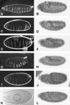
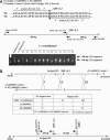
The Dpp signaling pathway is essential for many developmental processes in Drosophila and its activity is tightly regulated. To identify additional regulators of Dpp signaling, we conducted a genetic screen for maternal-effect suppressors of dpp haplo-insufficiency. We screened approximately 7000 EMS-mutagenized genomes and isolated and mapped seven independent dominant suppressors of dpp, Su(dpp), which were recovered as second-site mutations that resulted in viable flies in trans-heterozygous with dpp(H46), a dpp null allele. Most of the Su(dpp) mutants exhibited increased cell numbers of the amnioserosa, a cell type specified by the Dpp pathway, suggesting that these mutations may augment Dpp signaling activity. Here we report the unexpected identification of one of the Su(dpp) mutations as an allele of the eukaryotic translation initiation factor 4A (eIF4A). We show that Su(dpp)(YE9) maps to eIF4A and that this allele is associated with a substitution, arginine 321 to histidine, at a well-conserved amino acid and behaves genetically as a dominant-negative mutation. This result provides an intriguing link between a component of the translation machinery and Dpp signaling.
Figures


Similar articles
-
A novel function of Drosophila eIF4A as a negative regulator of Dpp/BMP signalling that mediates SMAD degradation.Nat Cell Biol. 2006 Dec;8(12):1407-14. doi: 10.1038/ncb1506. Epub 2006 Nov 19. Nat Cell Biol. 2006. PMID: 17115029 Free PMC article.
-
A genetic screen for modifiers of Drosophila decapentaplegic signaling identifies mutations in punt, Mothers against dpp and the BMP-7 homologue, 60A.Development. 1998 May;125(9):1759-68. doi: 10.1242/dev.125.9.1759. Development. 1998. PMID: 9521913
-
A genetic screen targeting the tumor necrosis factor/Eiger signaling pathway: identification of Drosophila TAB2 as a functionally conserved component.Genetics. 2005 Dec;171(4):1683-94. doi: 10.1534/genetics.105.045534. Epub 2005 Aug 3. Genetics. 2005. PMID: 16079232 Free PMC article.
-
Eukaryotic initiation factor 4A (eIF4A) during viral infections.Virus Genes. 2019 Jun;55(3):267-273. doi: 10.1007/s11262-019-01641-7. Epub 2019 Feb 22. Virus Genes. 2019. PMID: 30796742 Free PMC article. Review.
-
Dpp/BMP signaling in flies: from molecules to biology.Semin Cell Dev Biol. 2014 Aug;32:128-36. doi: 10.1016/j.semcdb.2014.04.036. Epub 2014 May 9. Semin Cell Dev Biol. 2014. PMID: 24813173 Review.
Cited by
-
A novel function of Drosophila eIF4A as a negative regulator of Dpp/BMP signalling that mediates SMAD degradation.Nat Cell Biol. 2006 Dec;8(12):1407-14. doi: 10.1038/ncb1506. Epub 2006 Nov 19. Nat Cell Biol. 2006. PMID: 17115029 Free PMC article.
-
Fatty Acid Synthase induced S6Kinase facilitates USP11-eIF4B complex formation for sustained oncogenic translation in DLBCL.Nat Commun. 2018 Feb 26;9(1):829. doi: 10.1038/s41467-018-03028-y. Nat Commun. 2018. PMID: 29483509 Free PMC article.
-
Loss of Paip1 causes translation reduction and induces apoptotic cell death through ISR activation and Xrp1.Cell Death Discov. 2023 Aug 5;9(1):288. doi: 10.1038/s41420-023-01587-8. Cell Death Discov. 2023. PMID: 37543696 Free PMC article.
References
-
- Brand, A. H., and N. Perrimon, 1993. Targeted gene expression as a means of altering cell fates and generating dominant phenotypes. Development 118: 401–415. - PubMed
-
- Brummel, T. J., V. Twombly, G. Marques, J. L. Wrana, S. J. Newfeld et al., 1994. Characterization and relationship of Dpp receptors encoded by the saxophone and thick veins genes in Drosophila. Cell 78: 251–261. - PubMed
-
- Campbell, G., and A. Tomlinson, 1999. Transducing the Dpp morphogen gradient in the wing of Drosophila: regulation of Dpp targets by brinker. Cell 96: 553–562. - PubMed
-
- Chen, Y., M. J. Riese, M. A. Killinger and F. M. Hoffmann, 1998. A genetic screen for modifiers of Drosophila decapentaplegic signaling identifies mutations in punt, Mothers against dpp and the BMP-7 homologue, 60A. Development 125: 1759–1768. - PubMed
Publication types
MeSH terms
Substances
Grants and funding
LinkOut - more resources
Full Text Sources
Molecular Biology Databases
Miscellaneous

