

Noninvasive biophotonic imaging for monitoring of catheter-associated urinary tract infections and therapy in mice
- PMID: 15972473
- PMCID: PMC1168591
- DOI: 10.1128/IAI.73.7.3878-3887.2005
Noninvasive biophotonic imaging for monitoring of catheter-associated urinary tract infections and therapy in mice
Abstract
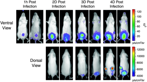
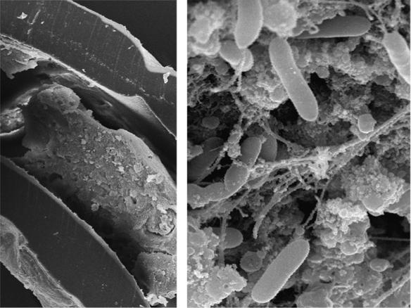
Urinary tract infections (UTIs) are among the most common bacterial infections acquired by humans, particularly in catheterized patients. A major problem with catheterization is the formation of bacterial biofilms on catheter material and the risk of developing persistent UTIs that are difficult to monitor and eradicate. To better understand the course of UTIs and allow more accurate studies of in vivo antibiotic efficacy, we developed a catheter-based biofilm infection model with mice, using bioluminescently engineered bacteria. Two important urinary tract pathogens, Pseudomonas aeruginosa and Proteus mirabilis, were made bioluminescent by stable insertion of a complete lux operon. Segments of catheter material (precolonized or postimplant infected) with either pathogen were placed transurethrally in the lumen of the bladder by using a metal stylet without surgical manipulation. The bioluminescent strains were sufficiently bright to be readily monitored from the outside of infected animals, using a low-light optical imaging system, including the ability to trace the ascending pattern of light-emitting bacteria through ureters to the kidneys. Placement of the catheter in the bladder not only resulted in the development of strong cystitis that persisted significantly longer than in mice challenged with bacterial suspensions alone but also required prolonged antibiotic treatment to reduce the level of infection. Treatment of infected mice for 4 days with ciprofloxacin at 30 mg/kg of body weight twice a day cured cystitis and renal infection in noncatheterized mice. Similarly, ciprofloxacin reduced the bacterial burden to undetectable levels in catheterized mice but did not inhibit rebound of the infection upon cessation of antibiotic therapy. This methodology easily allows spatial information to be monitored sequentially throughout the entire disease process, including ascending UTI, treatment efficacy, and relapse, all without exogenous sampling, which is not possible with conventional methods.
Figures


Similar articles
-
Clinical complications of urinary catheters caused by crystalline biofilms: something needs to be done.J Intern Med. 2014 Aug;276(2):120-9. doi: 10.1111/joim.12220. J Intern Med. 2014. PMID: 24635559 Review.
-
A high throughput, minimally invasive, ultrasound guided model for the study of catheter associated urinary tract infections and device encrustation in mice.J Urol. 2014 Dec;192(6):1856-63. doi: 10.1016/j.juro.2014.05.092. Epub 2014 May 24. J Urol. 2014. PMID: 24866596
-
Plasmid-mediated multiple antibiotic resistance in Proteus mirabilis isolated from patients with urinary tract infection.Med Sci Monit. 2004 Nov;10(11):CR598-602. Epub 2004 Oct 26. Med Sci Monit. 2004. PMID: 15507850
-
Antimicrobial activities against biofilm formed by Proteus mirabilis isolates from wound and urinary tract infections.Indian J Med Microbiol. 2012 Jan-Mar;30(1):76-80. doi: 10.4103/0255-0857.93044. Indian J Med Microbiol. 2012. PMID: 22361765
-
Bacterial characteristics of importance for recurrent urinary tract infections caused by Escherichia coli.Dan Med Bull. 2011 Apr;58(4):B4187. Dan Med Bull. 2011. PMID: 21466767 Review.
Cited by
-
Illuminating cellular physiology: recent developments.Sci Prog. 2007;90(Pt 2-3):129-60. doi: 10.3184/003685007X215986. Sci Prog. 2007. PMID: 17725230 Free PMC article. Review.
-
The Pathogenic Potential of Proteus mirabilis Is Enhanced by Other Uropathogens during Polymicrobial Urinary Tract Infection.Infect Immun. 2017 Jan 26;85(2):e00808-16. doi: 10.1128/IAI.00808-16. Print 2017 Feb. Infect Immun. 2017. PMID: 27895127 Free PMC article.
-
Complicated catheter-associated urinary tract infections due to Escherichia coli and Proteus mirabilis.Clin Microbiol Rev. 2008 Jan;21(1):26-59. doi: 10.1128/CMR.00019-07. Clin Microbiol Rev. 2008. PMID: 18202436 Free PMC article. Review.
-
Microbial Biofilms in Urinary Tract Infections and Prostatitis: Etiology, Pathogenicity, and Combating strategies.Pathogens. 2016 Nov 30;5(4):65. doi: 10.3390/pathogens5040065. Pathogens. 2016. PMID: 27916925 Free PMC article. Review.
-
Advances in experimental bladder models: bridging the gap between in vitro and in vivo approaches for investigating urinary tract infections.BMC Urol. 2024 Sep 23;24(1):206. doi: 10.1186/s12894-024-01590-w. BMC Urol. 2024. PMID: 39313789 Free PMC article. Review.
References
-
- Anderson, G. G., J. J. Palermo, J. D. Schilling, R. Roth, J. Heuser, and S. J. Hultgren. 2003. Intracellular bacterial biofilm-like pods in urinary tract infections. Science 301:105-107. - PubMed
-
- Berry, V., R. Page, J. Satterfield, C. Singley, R. Straub, and G. Woodnutt. 2000. Comparative efficacy of gemifloxacin in experimental models of pyelonephritis and wound infection. J. Antimicrob. Chemother. 45(Suppl. 1):87-93. - PubMed
-
- Contag, C. H., P. R. Contag, J. I. Mullins, S. D. Spilman, D. K. Stevenson, and D. A. Benaron. 1995. Photonic detection of bacterial pathogens in living hosts. Mol. Microbiol. 18:593-603. - PubMed
-
- Contag, P. R., I. N. Olomu, D. K. Stevenson, and C. H. Contag. 1998. Bioluminescent indicators in living mammals. Nat. Med. 4:245-247. - PubMed
-
- Davies, D. 2003. Understanding biofilm resistance to antibacterial agents. Nat. Rev. Drug Discov. 2:114-122. - PubMed
MeSH terms
LinkOut - more resources
Full Text Sources
Medical
