

Survival of Salmonella enterica serovar Typhimurium within late endosomal-lysosomal compartments of B lymphocytes is associated with the inability to use the vacuolar alternative major histocompatibility complex class I antigen-processing pathway
- PMID: 15972480
- PMCID: PMC1168566
- DOI: 10.1128/IAI.73.7.3937-3944.2005
Survival of Salmonella enterica serovar Typhimurium within late endosomal-lysosomal compartments of B lymphocytes is associated with the inability to use the vacuolar alternative major histocompatibility complex class I antigen-processing pathway
Abstract
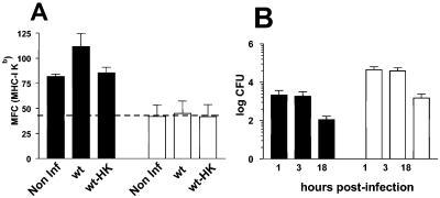
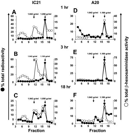
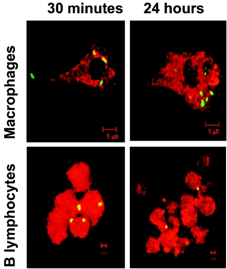
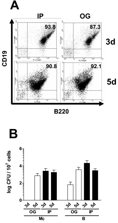
Gamma interferon (IFN-gamma)-activated macrophages use an alternative processing mechanism to present Salmonella antigens to CD8(+) T lymphocytes. This pathway involves processing of antigen in a vacuolar compartment followed by secretion and loading of antigenic peptides to major histocompatibility complex class I (MHC-I) molecules on macrophage cell surface and bystander cells. In this study, we have shown that B lymphocytes are not able to process Salmonella antigens using this alternative pathway. This is due to differences in Salmonella enterica serovar Typhimurium-containing vacuoles (SCV) when comparing late endosomal-lysosomal processing compartments in B lymphocytes to those in macrophages. The IFN-gamma-activated IC21 macrophage cell line and A-20 B-cell line were infected with live or dead Salmonella enterica serovar Typhimurium. The SCV in B cells were in a late endosomal-lysosomal compartment, whereas SCV in macrophages were remodeled to a non-characteristic late endosomal-lysosomal compartment over time. Despite the difference in SCV within macrophages and B lymphocytes, S. enterica serovar Typhimurium survives more efficiently within the IFN-gamma-activated B cells than in activated macrophage cell lines. Similar results were found during in vivo acute infection. We determined that a lack of remodeling of late endosomal-lysosomal compartments by live Salmonella infection in B lymphocytes is associated with the inability to use the alternative MHC-I antigen-processing pathway, providing a survival advantage to the bacterium. Our data also suggest that the B lymphocyte late endosome-lysosome environment allows the expression of Salmonella virulence mechanisms favoring B lymphocytes in addition to macrophages and dendritic cells as a reservoir during in vivo infection.
Figures

Similar articles
-
Processing of viable Salmonella typhimurium for presentation of a CD4 T cell epitope from the Salmonella invasion protein C (SipC).Eur J Immunol. 2002 Sep;32(9):2664-71. doi: 10.1002/1521-4141(200209)32:9<2664::AID-IMMU2664>3.0.CO;2-N. Eur J Immunol. 2002. PMID: 12207351
-
H2-M3 major histocompatibility complex class Ib-restricted CD8 T cells induced by Salmonella enterica serovar Typhimurium infection recognize proteins released by Salmonella serovar Typhimurium.Infect Immun. 2005 Dec;73(12):8002-8. doi: 10.1128/IAI.73.12.8002-8008.2005. Infect Immun. 2005. PMID: 16299293 Free PMC article.
-
Isolation and characterization of Salmonella typhimurium and Yersinia pseudotuberculosis-containing phagosomes from infected mouse macrophages: Y. pseudotuberculosis traffics to terminal lysosomes where they are degraded.Eur J Cell Biol. 1998 Sep;77(1):35-47. doi: 10.1016/S0171-9335(98)80100-3. Eur J Cell Biol. 1998. PMID: 9808287
-
Bacterial antigen delivery systems: phagocytic processing of bacterial antigens for MHC-I and MHC-II presentation to T cells.Behring Inst Mitt. 1997 Feb;(98):197-211. Behring Inst Mitt. 1997. PMID: 9382741 Review.
-
Eat in or Take out? Metabolism of Intracellular Salmonella enterica.Trends Microbiol. 2020 Aug;28(8):644-654. doi: 10.1016/j.tim.2020.03.005. Epub 2020 Apr 25. Trends Microbiol. 2020. PMID: 32345466 Review.
Cited by
-
MHC molecules and microbial antigen processing in phagosomes.Curr Opin Immunol. 2009 Feb;21(1):98-104. doi: 10.1016/j.coi.2009.01.001. Epub 2009 Feb 11. Curr Opin Immunol. 2009. PMID: 19217269 Free PMC article. Review.
-
SopB activates the Akt-YAP pathway to promote Salmonella survival within B cells.Virulence. 2018;9(1):1390-1402. doi: 10.1080/21505594.2018.1509664. Virulence. 2018. PMID: 30103648 Free PMC article.
-
Selective infection of antigen-specific B lymphocytes by Salmonella mediates bacterial survival and systemic spreading of infection.PLoS One. 2012;7(11):e50667. doi: 10.1371/journal.pone.0050667. Epub 2012 Nov 29. PLoS One. 2012. PMID: 23209805 Free PMC article.
-
Hemophagocytic macrophages harbor Salmonella enterica during persistent infection.PLoS Pathog. 2007 Dec;3(12):e193. doi: 10.1371/journal.ppat.0030193. PLoS Pathog. 2007. PMID: 18085823 Free PMC article.
-
Methylthioadenosine Suppresses Salmonella Virulence.Infect Immun. 2018 Aug 22;86(9):e00429-18. doi: 10.1128/IAI.00429-18. Print 2018 Sep. Infect Immun. 2018. PMID: 29866910 Free PMC article.
References
-
- Ackerman, A. L., and P. Cresswell. 2004. Cellular mechanisms governing cross-presentation of exogenous antigens. Nat. Immunol. 5:678-684. - PubMed
Publication types
MeSH terms
Substances
LinkOut - more resources
Full Text Sources
Research Materials
