

Nontoxic Shiga toxin derivatives from Escherichia coli possess adjuvant activity for the augmentation of antigen-specific immune responses via dendritic cell activation
- PMID: 15972497
- PMCID: PMC1168555
- DOI: 10.1128/IAI.73.7.4088-4097.2005
Nontoxic Shiga toxin derivatives from Escherichia coli possess adjuvant activity for the augmentation of antigen-specific immune responses via dendritic cell activation
Abstract
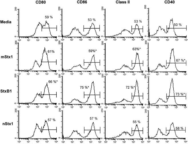
Shiga toxin (Stx) derivatives, such as the Stx1 B subunit (StxB1), which mediates toxin binding to the membrane, and mutant Stx1 (mStx1), which is a nontoxic doubly mutated Stx1 harboring amino acid substitutions in the A subunit, possess adjuvant activity via the activation of dendritic cells (DCs). Our results showed that StxB1 and mStx1, but not native Stx1 (nStx1), resulted in enhanced expression of CD86, CD40, and major histocompatibility complex (MHC) class II molecules and, to some extent, also enhanced the expression of CD80 on bone marrow-derived DCs. StxB1-treated DCs exhibited an increase in tumor necrosis factor alpha and interleukin-12 (IL-12) production, a stimulation of DO11.10 T-cell proliferation, and the production of both Th1 and Th2 cytokines, including gamma interferon (IFN-gamma), IL-4, IL-5, IL-6, and IL-10. When mice were given StxB1 subcutaneously, the levels of CD80, CD86, and CD40, as well as MHC class II expression by splenic DCs, were enhanced. The subcutaneous immunization of mice with ovalbumin (OVA) plus mStx1 or StxB1 induced high titers of OVA-specific immunoglobulin M (IgM), IgG1, and IgG2a in serum. OVA-specific CD4+ T cells isolated from mice immunized with OVA plus mStx1 or StxB1 produced IFN-gamma, IL-4, IL-5, IL-6, and IL-10, indicating that mStx1 and StxB1 elicit both Th1- and Th2-type responses. Importantly, mice immunized subcutaneously with tetanus toxoid plus mStx1 or StxB1 were protected from a lethal challenge with tetanus toxin. These results suggest that nontoxic Stx derivatives, including both StxB1 and mStx1, could be effective adjuvants for the induction of mixed Th-type CD4+ T-cell-mediated antigen-specific antibody responses via the activation of DCs.
Figures
Similar articles
-
Non-toxic Stx derivatives from Escherichia coli possess adjuvant activity for mucosal immunity.Vaccine. 2004 Sep 9;22(27-28):3751-61. doi: 10.1016/j.vaccine.2004.03.034. Vaccine. 2004. PMID: 15315856
-
Limiting dilution analysis of CD4 T-cell cytokine production in mice administered native versus polymerized ovalbumin: directed induction of T-helper type-1-like activation.Immunology. 1996 Jan;87(1):119-26. Immunology. 1996. PMID: 8666423 Free PMC article.
-
Activation of dendritic cells and induction of CD4(+) T cell differentiation by aluminum-containing adjuvants.Vaccine. 2007 Jun 6;25(23):4575-85. doi: 10.1016/j.vaccine.2007.03.045. Epub 2007 Apr 19. Vaccine. 2007. PMID: 17485153
-
Effects of cholera toxin on innate and adaptive immunity and its application as an immunomodulatory agent.J Leukoc Biol. 2004 May;75(5):756-63. doi: 10.1189/jlb.1103534. Epub 2004 Jan 2. J Leukoc Biol. 2004. PMID: 14704372 Review.
-
Mucosal immunity: regulation by helper T cells and a novel method for detection.J Biotechnol. 1996 Jan 26;44(1-3):209-16. doi: 10.1016/0168-1656(95)00095-X. J Biotechnol. 1996. PMID: 8717406 Review.
Cited by
-
Cationic-nanogel nasal vaccine containing the ectodomain of RSV-small hydrophobic protein induces protective immunity in rodents.NPJ Vaccines. 2023 Jul 24;8(1):106. doi: 10.1038/s41541-023-00700-3. NPJ Vaccines. 2023. PMID: 37488116 Free PMC article.
-
Analyzing of expression of novel polypeptide complexes consisting of Shiga toxin B subunit and Adherence Fimbriae of Escherichia coli based on in silico modeling.J Mol Model. 2012 Sep;18(9):4131-9. doi: 10.1007/s00894-012-1414-3. Epub 2012 Apr 24. J Mol Model. 2012. PMID: 22527278
-
Shiga toxin (Stx)1B and Stx2B induce von Willebrand factor secretion from human umbilical vein endothelial cells through different signaling pathways.Blood. 2011 Sep 22;118(12):3392-8. doi: 10.1182/blood-2011-06-363648. Epub 2011 Aug 3. Blood. 2011. PMID: 21816831 Free PMC article.
-
The Role of Escherichia coli Shiga Toxins in STEC Colonization of Cattle.Toxins (Basel). 2020 Sep 21;12(9):607. doi: 10.3390/toxins12090607. Toxins (Basel). 2020. PMID: 32967277 Free PMC article. Review.
-
The mucosal immune system: From dentistry to vaccine development.Proc Jpn Acad Ser B Phys Biol Sci. 2015;91(8):423-39. doi: 10.2183/pjab.91.423. Proc Jpn Acad Ser B Phys Biol Sci. 2015. PMID: 26460320 Free PMC article. Review.
References
-
- Agren, L. C., L. Ekman, B. Lowenadler, and N. Y. Lycke. 1997. Genetically engineered nontoxic vaccine adjuvant that combines B cell targeting with immunomodulation by cholera toxin A1 subunit. J. Immunol. 158:3936-3946. - PubMed
-
- Austin, P. R., and C. J. Hovde. 1995. Purification of recombinant Shiga-like toxin type I B subunit. Protein Expr. Purif. 6:771-779. - PubMed
-
- Birnabaum, S., and M. Pinto. 1976. Local and systemic opsonic adherent, hemagglutinating and rosette forming activity in mice induced by respiratory immunization with sheep red blood cells. Z. Immunitatsforsch. Exp. Klin. Immunol. 151:69-77. - PubMed
-
- Bluestone, J. A. 1995. New perspectives of CD28-B7-mediated T cell costimulation. Immunity 2:555-559. - PubMed
-
- Byun, Y., M. Ohmura, K. Fujihashi, S. Yamamoto, J. McGhee, S. Udaka, H. Kiyono, Y. Takeda, T. Kohsaka, and Y. Yuki. 2001. Nasal immunization with E. coli verotoxin 1 (VT1)-B subunit and a nontoxic mutant of cholera toxin elicits serum neutralizing antibodies. Vaccine 19:2061-2070. - PubMed
Publication types
MeSH terms
Substances
LinkOut - more resources
Full Text Sources
Research Materials
