. 2005 Jul;73(7):4180-9.
doi: 10.1128/IAI.73.7.4180-4189.2005.

Impact of Helicobacter pylori virulence factors and compounds on activation and maturation of human dendritic cells

Affiliations

Impact of Helicobacter pylori virulence factors and compounds on activation and maturation of human dendritic cells

Katharina Kranzer et al. Infect Immun. 2005 Jul.

Abstract

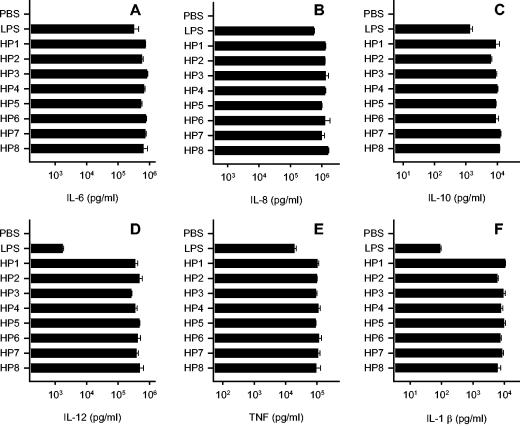
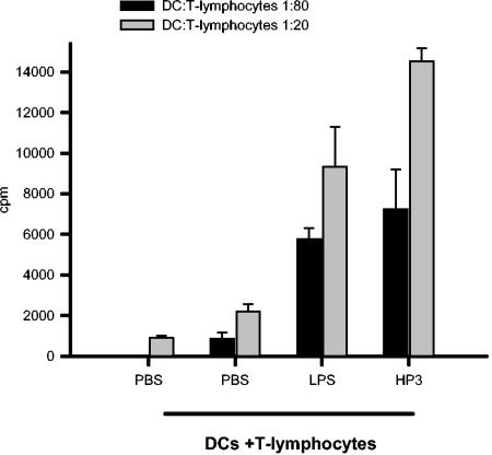
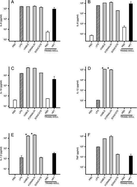
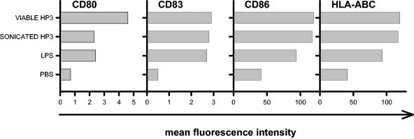
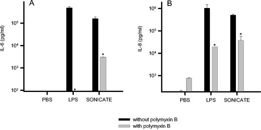
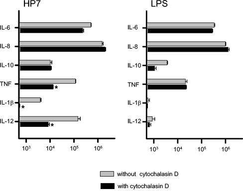
Recently, we and others have shown that Helicobacter pylori induces dendritic cell (DC) activation and maturation. However, the impact of virulence factors on the interplay between DCs and H. pylori remains elusive. Therefore, we investigated the contribution of cag pathogenicity island (PAI) and VacA status on cytokine release and up-regulation of costimulatory molecules in H. pylori-treated DCs. In addition, to characterize the stimulatory capacity of H. pylori compounds in more detail, we studied the effect of formalin-inactivated and sonicated H. pylori, as well as secreted H. pylori molecules, on DCs. Incubation of DCs with viable or formalin-inactivated H. pylori induced comparable secretion of interleukin-6 (IL-6), IL-8, IL-10, IL-12, IL-1beta, and tumor necrosis factor (TNF). In contrast, IL-12 and IL-1beta release was significantly reduced in DCs treated with sonicated bacteria and secreted bacterial molecules. Treatment of sonicated H. pylori preparations with polymyxin B resulted in a significant reduction of IL-8 and IL-6 secretion, suggesting that H. pylori-derived lipopolysaccharide at least partially contributes to activation of immature DCs. In addition, the capacity of H. pylori-pulsed DCs to activate allogeneic T cells was not affected by cag PAI and VacA. Pretreatment of DC with cytochalasin D significantly inhibited secretion of IL-12, IL-1beta, and TNF, indicating that phagocytosis of H. pylori contributes to maximal activation of DCs. Taken together, our results suggest that DC activation and maturation, as well as DC-mediated T-cell activation, are independent of the cag PAI and VacA status of H. pylori.


Figures

FIG. 1.
FIG. 1.
Cytokines released by DCs stimulated with H. pylori. A total of 106 DCs/ml were pulsed with different H. pylori strains (MOI, 10) for 24 h. Controls included DCs stimulated with PBS (negative control) and E. coli-LPS at a concentration of 100 ng/ml (positive control). The concentrations of cytokines secreted into the medium were assayed by ELISA: IL-6 (A), IL-8 (B), IL-10 (C), IL-12 (D), TNF (E), and IL-1β (F). One representative experiment out of five is shown. Data represent means ± SD.
FIG. 2.
FIG. 2.
Effects of different H. pylori strains on DC maturation. A total of 106 DCs/ml were stimulated with different H. pylori strains (MOI, 10) for 72 h. Controls included DCs treated with PBS (negative control) and E. coli-LPS at a concentration of 100 ng/ml (positive control). CD80, CD83, CD86, and HLA-ABC expression was determined by flow cytometry and is shown as isotype-corrected MFI. One representative experiment out of five is shown.
FIG. 3.
FIG. 3.
Stimulatory ability of DC in the MLR assay. DCs were treated with H. pylori, E. coli-LPS (positive control), and PBS (negative control) for 24 h. DCs were harvested and washed twice with PBS before being cocultured with allogeneic T cells (5 × 10⁴/well) in 96-well tissue plates at a DC/T-cell ratio of 1:20 or 1:80. Proliferation of T cells was measured by uptake of [3H]thymidine on day 6. One representative experiment out of four is shown. Data represent means ± SD.
FIG. 4.
FIG. 4.
Influence of soluble and integral H. pylori (HP7) factors on cytokine production. A total of 106 DCs/ml were stimulated for 24 h with viable, formalin-inactivated, heat-inactivated H. pylori, H. pylori sonicate (grey bars), and soluble bacterial molecules from trans-well cultures (black bars). Controls included DCs treated with PBS (open bars) directly or via trans-well cultures (negative controls) and E. coli-LPS (shaded bars) at a concentration of 100 ng/ml (positive control). Cytokine secretion of IL-6 (A), IL-8 (B), IL-10 (C), IL-12 (D), IL-1β (E), and TNF (F) was determined by ELISA. One representative experiment out of four is shown. Data represent means ± SD. *, P < 0.05.
FIG. 5.
FIG. 5.
Effect of H. pylori components on DC maturation. A total of 106 DCs/ml were stimulated with viable H. pylori strains or H. pylori sonicate (100 μg/ml) for 72 h. Controls included DCs treated with PBS (negative control) and E. coli LPS at a concentration of 100 ng/ml (positive control). CD80, CD83, CD86, and HLA-ABC expression was determined by flow cytometry and is shown as isotype-corrected MFI. One representative experiment out of four is shown.
FIG. 6.
FIG. 6.
Effect of polymyxin B treatment on H. pylori sonicate-induced activation of DCs. A total of 106 DCs/ml were stimulated for 24 h with H. pylori sonicate (100 μg/ml) or E. coli LPS (100 ng/ml) in the presence of either medium alone or the LPS antagonist polymyxin B (200 μg/ml). Cytokine concentrations of IL-6 (A) and IL-8 (B) were determined from culture supernatants by ELISA. One representative experiment out of five is shown. Data represent means ± SD. *, P < 0.05.
FIG. 7.
FIG. 7.
Reduction of cytokine production by inhibition of bacterial internalization. A total of 106 DCs/ml were either preincubated with medium alone or cytochalasin D (500 ng/ml) before stimulation with H. pylori or E. coli LPS (100 ng/ml). The culture supernatants were collected after 24 h, and cytokine concentration was determined by ELISA. One representative experiment out of four is shown. Data represent means ± SD. *, P < 0.05.

Similar articles

Cited by

References

    1. Akanuma, M., S. Maeda, K. Ogura, Y. Mitsuno, Y. Hirata, T. Ikenoue, M. Otsuka, T. Watanabe, Y. Yamaji, H. Yoshida, T. Kawabe, Y. Shiratori, and M. Omata. 2002. The evaluation of putative virulence factors of Helicobacter pylori for gastroduodenal disease by use of a short-term Mongolian gerbil infection model. J. Infect. Dis. 185:341-347. - PubMed
    1. Allen, L. A. 2003. Mechanisms of pathogenesis: evasion of killing by polymorphonuclear leukocytes. Microbes Infect. 5:1329-1335. - PubMed
    1. Allen, L. A., L. S. Schlesinger, and B. Kang. 2000. Virulent strains of Helicobacter pylori demonstrate delayed phagocytosis and stimulate homotypic phagosome fusion in macrophages. J. Exp. Med. 191:115-128. - PMC - PubMed
    1. Amieva, M. R., R. Vogelmann, A. Covacci, L. S. Tompkins, W. J. Nelson, and S. Falkow. 2003. Disruption of the epithelial apical-junctional complex by Helicobacter pylori CagA. Science 300:1430-1434. - PMC - PubMed
    1. Asaka, M., M. Kudo, M. Kato, T. Kimura, T. Meguro, S. Mitani, T. Miyazaki, and K. Inoue. 1994. The role of Helicobacter pylori infection in the pathogenesis of gastritis. J. Gastroenterol. 29(Suppl. 7):100-104. - PubMed

Publication types

LinkOut - more resources