. 2005 Jul 1;175(1):196-200.
doi: 10.4049/jimmunol.175.1.196.

CD8alpha+ dendritic cells selectively present MHC class I-restricted noncytolytic viral and intracellular bacterial antigens in vivo

Affiliations

CD8alpha+ dendritic cells selectively present MHC class I-restricted noncytolytic viral and intracellular bacterial antigens in vivo

Gabrielle T Belz et al. J Immunol. .

Abstract

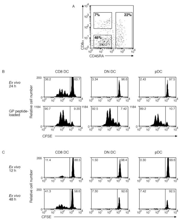
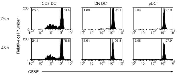
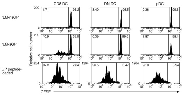
CD8alpha(+) dendritic cells (DCs) have been shown to be the principal DC subset involved in priming MHC class I-restricted CTL immunity to a variety of cytolytic viruses, including HSV type 1, influenza, and vaccinia virus. Whether priming of CTLs by CD8alpha(+) DCs is limited to cytolytic viruses, which may provide dead cellular material for this DC subset, or whether these DCs selectively present intracellular Ags, is unknown. To address this question, we examined Ag presentation to a noncytolytic virus, lymphocytic choriomeningitis virus, and to an intracellular bacterium, Listeria monocytogenes. We show that regardless of the type of intracellular infection, CD8alpha(+) DCs are the principal DC subset that initiate CD8(+) T cell immunity.


Conflict of interest statement

Disclosures

The authors have no financial conflict of interest.

Figures

FIGURE 1
FIGURE 1
CD8 DCs are the only subset that activates naive CD8+ T cells after i.v. infection with LCMV virus. A, Sorting gates for DCs enriched from spleen at various time points after infection. Cells were enriched for DCs by magnetic depletion of cells staining for CD3, Thy-1, CD19, anti-Gr-1, and erythrocytes. Enriched cells were then stained with anti-CD11c, anti-CD8α, and anti-CD45RA. Rectangles show sorting gates for CD8α+CD45RA (CD8), CD45RA+ (pDC), and CD8αCD45RA DC (DN DCs). B, Mice were infected with LCMV by i.v. inoculation 24 h after infection; DCs were enriched from spleen and sorted by flow cytometry into CD8 DC, DN DC, or pDC before culturing 2.5 × 10⁴ cells with 5 × 10⁴ CFSE-labeled glycoprotein-specific CD8+ T cells. Some DCs were coated with 0.1 μM glycoprotein peptide for 1 h at 37°C and washed three times before coculture with CFSE-labeled glycoprotein-specific CD8+ T cells (lower panel). Proliferation was analyzed at 60 h of culture. The percentage of proliferating cells for each culture is indicated in the top left corner of each histogram (C). At 12 and 48 h after infection, DCs were enriched from spleen of infected mice and responses were analyzed as in B. Each time point was performed twice with similar results. A 6-h time point was also examined in a single experiment.
FIGURE 2
FIGURE 2
CD8 DCs, but not other DCs, activate naive LCMV-specific CD8+ T cells after i.p. infection with virus. Mice were infected with LCMV by i.p. injection. One or 2 days after infection, DCs were enriched from spleen and then sorted into CD8 DC, DN DC, and pDC before culturing 2.5 × 10⁴ cells with 5 × 10⁴ CFSE-labeled glycoprotein-specific CD8+ T cells. Proliferation was analyzed at 60 h of culture. The percentage of proliferating cells for each culture is indicated in the top left corner of each histogram. This experiment was performed twice with similar results.
FIGURE 3
FIGURE 3
Only CD8 DCs present rLM expressing the glycoprotein Ag. Mice were infected with nonsecreted (upper panel) or secreted (middle panel) forms of the rLM glycoprotein (29) by i.v. injection. DCs were enriched from the spleen 24 h after infection and sorted by flow cytomety into CD8 DC, DN DC, or pDC. Purified DC subsets (2.5 × 10⁴ DC) were then cocultured with 5 × 10⁴ CFSE-labeled glycoprotein-specific CD8+ T cells for 60 h. Twenty-four hour infected DCs from each subset were coated with 0.1 μM glycoprotein peptide for 1 h at 37°C and washed three times before coculture with CFSE-labeled glycoprotein-specific CD8+ T cells (lower panel). The percentage of proliferating cells for each culture is indicated in the top left corner of each histogram. Each experiment was performed twice with similar results. Ag presentation was also examined 12 and 18 h after infection with secreted and nonsecreted forms of rLM glycoprotein (data not shown; each experiment was performed once). No proliferation of naive glycoprotein-specific CD8+ T cells was detected at either of these time points.

Similar articles

Cited by

References

    1. Jung S, Unutmaz D, Wong P, Sano G, De los Santos K, Sparwasser T, Wu S, Vuthoori S, Ko K, Zavala F, et al. In vivo depletion of CD11c+ dendritic cells abrogates priming of CD8+ T cells by exogenous cell-associated antigens. Immunity. 2002;17:211–220.x. - PMC - PubMed
    1. Vremec D, Pooley J, Hochrein H, Wu L, Shortman K. CD4 and CD8 expression by dendritic cell subtypes in mouse thymus and spleen. J Immunol. 2000;164:2978–2986. - PubMed
    1. Vremec D, Zorbas M, Scollay R, Saunders DJ, Ardavin CF, Wu L, Shortman K. The surface phenotype of dendritic cells purified from mouse thymus and spleen: investigation of the CD8 expression by a subpopulation of dendritic cells. J Exp Med. 1992;176:47–58. - PMC - PubMed
    1. O’Keeffe M, Hochrein H, Vremec D, Caminschi I, Miller JL, Anders EM, Wu L, Lahoud MH, Henri S, Scott B, et al. Mouse plasmacytoid cells: long-lived cells, heterogeneous in surface phenotype and function, that differentiate into CD8+ dendritic cells only after microbial stimulus. J Exp Med. 2002;196:1307–1319. - PMC - PubMed
    1. Shortman K, Liu YJ. Mouse and human dendritic cell subtypes. Nat Rev Immunol. 2002;2:151–161. - PubMed

Publication types

MeSH terms