. 2005 Jul 1;175(1):413-20.
doi: 10.4049/jimmunol.175.1.413.

Novel innate immune functions for galectin-1: galectin-1 inhibits cell fusion by Nipah virus envelope glycoproteins and augments dendritic cell secretion of proinflammatory cytokines

Affiliations

Novel innate immune functions for galectin-1: galectin-1 inhibits cell fusion by Nipah virus envelope glycoproteins and augments dendritic cell secretion of proinflammatory cytokines

Ernest L Levroney et al. J Immunol. .

Abstract

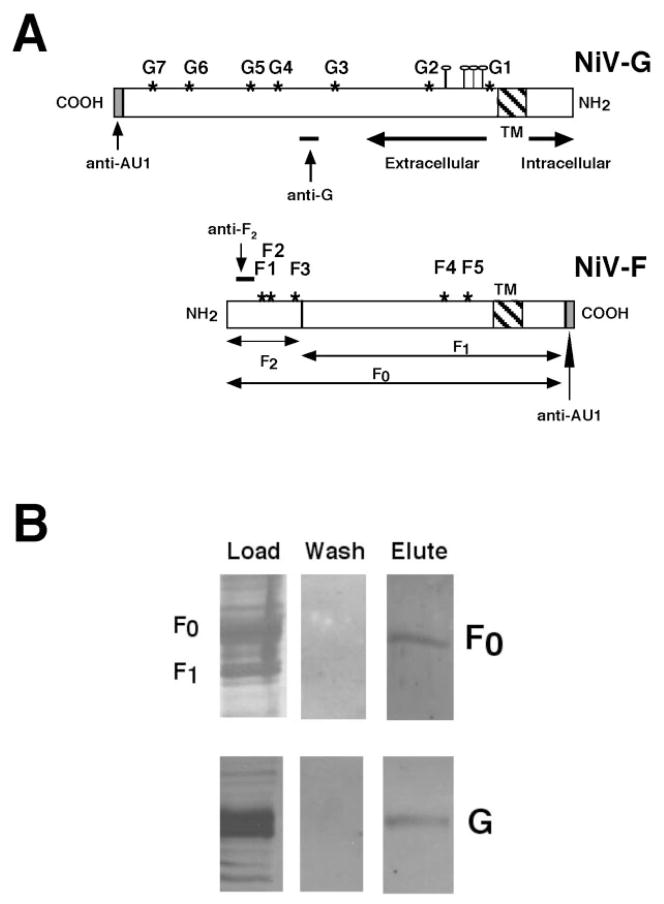
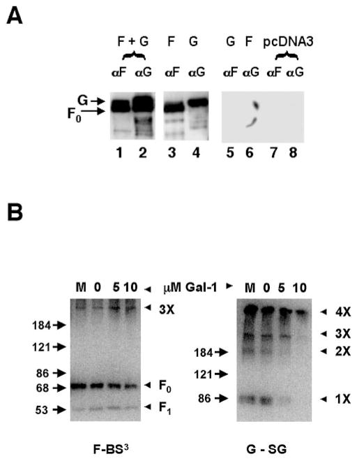
Galectin-1 (gal-1), an endogenous lectin secreted by a variety of cell types, has pleiotropic immunomodulatory functions, including regulation of lymphocyte survival and cytokine secretion in autoimmune, transplant disease, and parasitic infection models. However, the role of gal-1 in viral infections is unknown. Nipah virus (NiV) is an emerging pathogen that causes severe, often fatal, febrile encephalitis. The primary targets of NiV are endothelial cells. NiV infection of endothelial cells results in cell-cell fusion and syncytia formation triggered by the fusion (F) and attachment (G) envelope glycoproteins of NiV that bear glycan structures recognized by gal-1. In the present study, we report that NiV envelope-mediated cell-cell fusion is blocked by gal-1. This inhibition is specific to the Paramyxoviridae family because gal-1 did not inhibit fusion triggered by envelope glycoproteins of other viruses, including two retroviruses and a pox virus, but inhibited fusion triggered by envelope glycoproteins of the related Hendra virus and another paramyxovirus. The physiologic dimeric form of gal-1 is required for fusion inhibition because a monomeric gal-1 mutant had no inhibitory effect on cell fusion. gal-1 binds to specific N-glycans on NiV glycoproteins and aberrantly oligomerizes NiV-F and NiV-G, indicating a mechanism for fusion inhibition. gal-1 also increases dendritic cell production of proinflammatory cytokines such as IL-6, known to be protective in the setting of other viral diseases such as Ebola infections. Thus, gal-1 may have direct antiviral effects and may also augment the innate immune response against this emerging pathogen.


Conflict of interest statement

Disclosures

The authors have no financial conflict of interest.

Figures

FIGURE 1
FIGURE 1
Schematic of NiV-G and NiV-F envelope glycoproteins. A, NiV-G is a type II transmembrane protein. NiV-F (F0) is a type I transmembrane protein cleaved into F1 and F2 by a host cell protease. *, Potential N-linked glycosylation sites, F1 to F5 and G1 to G7. ⫯, Potential NiV-G O-linked glycosylation sites. Epitopes of NiV-F and NiV-G antipeptide Abs are indicated. Codon-optimized NiV-G and NiV-F were AU1 epitope tagged at the indicated termini. B, NiV-F and NiV-G bind gal-1. Extracts of cell membranes from 293T cells expressing either NiV-F or NiV-G were applied to a gal-1 affinity column. Bound glycoproteins were eluted with β-lactose and detected with anti-AU1 mAb. No glycoproteins were eluted with sucrose wash.
FIGURE 2
FIGURE 2
gal-1 blocks NiV envelope-mediated fusion. A, gal-1 inhibits NiV-F- and NiV-G-mediated fusion. Vero cells expressing NiV-F and NiV-G were cultured for 16 h in medium with gal-1 (20 μM in 0.32 mM DTT) or buffer control. gal-1 has unpaired cysteines in the binding pocket that can form intra- or intermolecular disulfide binds in the absence of saccharide ligands, which can lead to inactivation of the binding site. Thus, the buffer control includes the same concentration of DTT, a reducing agent, which prevents gal-1 inactivation. To demonstrate the specificity of gal-1 inhibition, rabbit anti-gal-1 serum (in increasing amounts) or preimmune serum (IgG) were added simultaneously with gal-1. Fusion was quantified as described in Materials and Methods. Values of p were calculated using the Student t test (unpaired samples with unequal variance). Mean ± SD of one representative experiment of three is presented. Note that anti-gal-1 sera alone did not inhibit NiV-G/F-mediated fusion. B, Specificity of gal-1 inhibition for Paramyxoviridae family: inhibition was observed only for fusion mediated by Hendra virus (HeV) glycoproteins and human parainfluenza virus type III (HPIV III), whereas no significant inhibition was seen for fusion mediated by glycoproteins from human T lymphotrophic virus II (HTLV II), murine leukemia virus (MLV), or vaccinia (WR strain). Mean ± SE of two determinations. C, Lactose reversal of gal-1 inhibition. A total of 10 mM lactose, a saccharide ligand for gal-1, or 10 mM sucrose, which does not bind gal-1, was added to fusion assays concurrently with gal-1. Fusion was quantified as above. Mean ± SE of two determinations.
FIGURE 3
FIGURE 3
Dimeric gal-1 is required to block NiV-F/G-mediated fusion. A, Monomeric (N-Gal-1) or dimeric gal-1 was added to Vero cells transfected with NiV-F and NiV-G. Fusion was quantified as in Fig. 2. Note that monomeric gal-1 had no effect on fusion even up to 25 μM. Mean ± SD of one representative experiment of three is presented. B, N-Gal-1 binds to T cells in a lactose inhibitable fashion. Increasing concentrations of biotinylated monomeric gal-1 were added to T cells. After binding for 30 min at 4°C, cells were washed once in 1× PBS, and bound gal-1 was detected by addition of FITC-conjugated streptavidin and flow cytometry. The highest amount of N-Gal-1 binding observed was reversed by the addition of 0.1 mM lactose. MFI, mean fluorescence intensity. C, N-Gal-1 is monomeric. gal-1 or N-Gal-1 was added to CEM T cells for 30 or 180 min, and the cells examined by phase-contrast microscopy. Dimeric gal-1 agglutinated T cells (arrows) due to the ability to bind carbohydrate ligands on adjacent cells. N-Gal-1 was unable to agglutinate T cells, indicating that N-Gal-1 is monomeric at this concentration.
FIGURE 4
FIGURE 4
gal-1 binds to a specific N-glycan site in NiV-F and NiV-G. A, F1, F2, F4, and F5 mutants are as sensitive to gal-1 inhibition as wt NiV-F. Vero cells transfected with NiV-G and wt NiV-F or the indicated NiV-F mutants (F1, F2, F4, or F5) were cultured with indicated concentrations of gal-1 and fusogenicity determined as in Fig. 1. Ten fields were counted for each mutant per condition. The number of nuclei in syncytia per field with no gal-1 treatment is normalized to 100% (fusogenic index). Mean ± SD of one representative experiment of three is presented. B, NiV-F3 glycan mutant is resistant to gal-1 inhibition of fusion. For clarity, results are shown only for wt NiV-F and F3 only. The experiment was performed identically as described in A. C, The NiV-F3 mutant reduces gal-1 binding. gal-1 (20 μM) was added to 293T cells expressing equivalent amounts of NiV-F or NiV-F3 protein, cell lysates were immunoprecipitated with rabbit anti-gal-1 antiserum, and NiV-F and NiV-F3 were detected with anti-AU1. D, gal-1 binding to NiV-F is dependent on lectin-glycan interaction. DMNJ (2 mM), which blocks the processing required for formation of complex/hybrid N-glycans, was added to 293T cells expressing NiV-F for 48 h. gal-1 (20 μM) was added to the cells, the cells were lysed, and the lysates were immunoprecipitated with an anti-gal-1 Ab. Precipitated proteins were detected with anti-AU1. CL indicates the input cell lysate while IP indicated precipitated protein. Note that DMNJ treatment itself did not affect levels of NiV-F.
FIGURE 5
FIGURE 5
gal-1 modulates the oligomeric state of NiV-F and NiV-G. A, Association of NiV-F and NiV-G. Extracts of 293T cells expressing NiV-F and NiV-G (F + G), or NiV-F or NiV-G alone, were immunoprecipitated with anti-F peptide or anti-G peptide antisera. Precipitated proteins were detected with anti-AU1 to detect both NiV-F and NiV-G. Anti-F and anti-G coimmunoprecipitated NiV-G and NiV-F, respectively (lanes 1 and 2). When NiV-F and NiV-G were expressed alone, anti-F and anti-G precipitated only the relevant glycoproteins (lanes 3–6). No precipitated protein was seen in cells transfected with plasmid alone (lanes 7 and 8). B, gal-1 modulates oligomerization of NiV-F and NiV-G. gal-1 was added to 293T cells expressing NiV-F or NiV-G. Cell surface proteins were cross-linked using membrane impermeant cross-linkers (BS3 for NiV-F and sulfo-GMBS (SG) for NiV-G), and NiV-F and NiV-G were detected with anti-AU1. Uncleaved NiV-F monomer (F0) and the estimated NiV-F trimer (3×) are indicated. Molecular mass markers are shown. Molecular mass estimates >180 kDa are not precise, but for NiV-G, the progressive laddering pattern suggests that monomers (1×), dimers (2×), trimers (3×), and tetramers (4×) are formed. M, 10 mM monomeric N-Gal-1, does not enhance oligomerization.
FIGURE 6
FIGURE 6
gal-1 induces DC secretion of proinflammatory cytokines. Human PBMCs were cultured for 5 days with IL-4 (100 ng/ml) + GM-CSF (50 ng/ml). At day 5, 20 μM gal-1 (with or without 100 mM lactose) or N-Gal-1 were added, and the culture was continued for an additional 48 h. Cytokine secretion was measured in tissue culture supernatant by cytometric bead array. One experiment of three is shown. *, Lower limit of the linear range.

Similar articles

Cited by

References

    1. Wong KT, Shieh WJ, Kumar S, Norain K, Abdullah W, Guarner J, Goldsmith CS, Chua KB, Lam SK, Tan CT, et al. Nipah virus infection: pathology and pathogenesis of an emerging paramyxoviral zoonosis. Am J Pathol. 2002;161:2153–2167. - PMC - PubMed
    1. Chua KB, Bellini WJ, Rota PA, Harcourt BH, Tamin A, Lam SK, Ksiazek TG, Rollin PE, Zaki SR, Shieh W, et al. Nipah virus: a recently emergent deadly paramyxovirus. Science. 2000;288:1432–1435. - PubMed
    1. Lam SK. Nipah virus: a potential agent of bioterrorism? Antiviral Res. 2003;57:113–119. - PubMed
    1. Nipah virus outbreak(s) in Bangladesh, January–April 2004. Wkly Epidemiol Rec. 2004;79:168. - PubMed
    1. Mackenzie JS, Chua KB, Daniels PW, Eaton BT, Field HE, Hall RA, Halpin K, Johansen CA, Kirkland PD, Lam SK, et al. Emerging viral diseases of Southeast Asia and the Western Pacific. Emerg Infect Dis. 2001;7(3 Suppl):497–504. - PMC - PubMed

Publication types

MeSH terms