Comparative Study
. 2005 Jul 5;102(27):9559-64.
doi: 10.1073/pnas.0409553102. Epub 2005 Jun 22.

Nonlinear protein degradation and the function of genetic circuits

Affiliations
Comparative Study

Nonlinear protein degradation and the function of genetic circuits

Nicolas E Buchler et al. Proc Natl Acad Sci U S A. .

Abstract

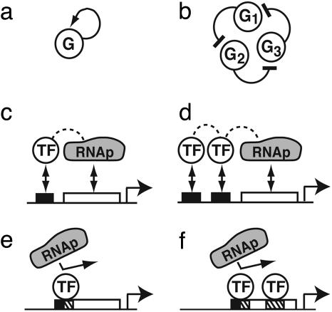
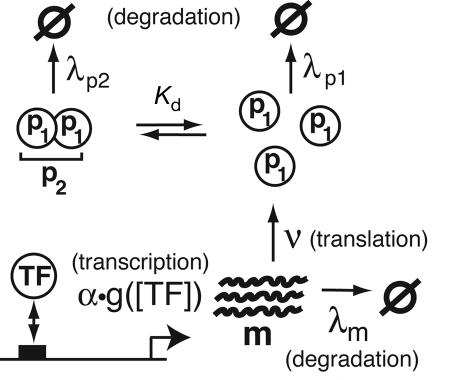
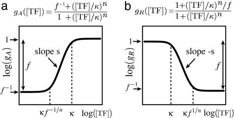
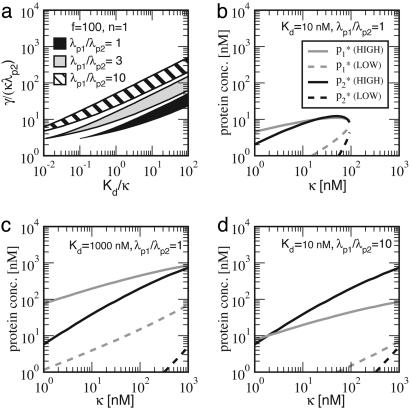
The functions of most genetic circuits require a sufficient degree of cooperativity in the circuit components. Although mechanisms of cooperativity have been studied most extensively in the context of transcriptional initiation control, cooperativity from other processes involved in the operation of the circuits can also play important roles. In this work, we examine a simple kinetic source of cooperativity stemming from the nonlinear degradation of multimeric proteins. Ample experimental evidence suggests that protein subunits can degrade less rapidly when associated in multimeric complexes, an effect we refer to as "cooperative stability." For dimeric transcription factors, this effect leads to a concentration-dependence in the degradation rate because monomers, which are predominant at low concentrations, will be more rapidly degraded. Thus, cooperative stability can effectively widen the accessible range of protein levels in vivo. Through theoretical analysis of two exemplary genetic circuits in bacteria, we show that such an increased range is important for the robust operation of genetic circuits as well as their evolvability. Our calculations demonstrate that a few-fold difference between the degradation rate of monomers and dimers can already enhance the function of these circuits substantially. We discuss molecular mechanisms of cooperative stability and their occurrence in natural or engineered systems. Our results suggest that cooperative stability needs to be considered explicitly and characterized quantitatively in any systematic experimental or theoretical study of gene circuits.


Figures

Fig. 1.
Fig. 1.
Two simple genetic circuits. (a and b) The circuits shown are capable of bistability (a) and oscillation (b). Genetic circuits consist of genes (drawn as circles) that regulate the transcriptional activity of one another. This regulation can be activating (arrow) or repressive (blunt line). (cf) Exemplary cis-regulatory architectures in bacteria by using one (c) or two (d) operator sites for activation and one (e) or two (f) operator sites for repression. The core promoter to which RNA polymerase (RNAp) binds and the operator sites to which the TFs bind are drawn as open or black boxes, respectively. The dashed lines depict cooperative interaction between regulatory proteins, whereas overlapping operators (indicated by hatched boxes) denote repression mediated through excluded volume interaction.
Fig. 2.
Fig. 2.
Schematic of the basic parameters involved in transcription, translation, degradation, and dimerization. Transcription is governed by the transcription rate α·g([TF]), where α is the mRNA synthesis rate at full activation. Each mRNA is translated into protein monomer at a rate ν and degraded at a rate λm. The cellular concentrations of monomers (p1) and dimers (p2) are related by the dimer dissociation constant Kd. The protein degradation rate can be different for monomers (λp1) and dimers (λp2).
Fig. 3.
Fig. 3.
Log–log plot of the relative promoter activity g([TF]) vs. the TF concentration [TF] for activation (a) and repression (b). The general expression for the promoter activity function is written above each plot. The peak activity of a promoter is defined to be 1, the fold-change between LOW and HIGH plateaus is described by f, and the DNA-binding dissociation constant of a TF for its operator (κ) is the concentration that separates the HIGH plateau from the transition region. The log–log slope (s) of the transition region [referred to as “sensitivity” or “gain” in the literature (10)] quantifies the degree of cooperativity in transcriptional control. It is determined by the Hill coefficient n and the maximum fold-change f, with maximum s approaching n for large f (see Supporting Text). Both a and b are approximations to promoter activity functions derived from the detailed thermodynamic treatment of transcriptional initiation (see Supporting Text and refs. –14).
Fig. 4.
Fig. 4.
Quantitative characteristics of the bistable circuit with a single operator promoter (n = 1) and a strong activator (f = 100). (a) Regime of bistability in the parameter space for the circuit with linear degradation (λp1p2 = 1) and with cooperative stability (λp1p2 > 1). The axes show combinations of the parameters that are both useful for discussion and natural in the quantitative description (see Supporting Text). (b and c) For linear degradation, the steady-state monomer (gray) and dimer (black) concentrations (i.e., formula image and formula image, respectively) are plotted for different values of κ, with Kd = 10 nM (b) and 1,000 nM (c). For each choice of Kd and κ, γ is chosen such that the system is in the middle of the bistable regime, i.e., the black band in a. For both formula image and formula image, the solid curve is the concentration in the HIGH bistable state and the dashed curve is the concentration in the LOW bistable state. (d) Same plot as b for the circuit with cooperative stability (λp1p2 = 10).

Similar articles

Cited by

References

    1. Gottesman, S. & Maurizi, M. R. (1992) Microbiol. Rev. 56 592-621. - PMC - PubMed
    1. Jenal, U. & Hengge-Aronis, R. (2003) Curr. Opin. Microbiol. 6 163-172. - PubMed
    1. Gonzalez, M. & Woodgate, R. (2002) BioEssays 24 141-148. - PubMed
    1. Johnson, P. R., Swanson, R., Rakhilina, L. & Hochstrasser, M. (1998) Cell 94 217-227. - PubMed
    1. Parsell, D. A. & Sauer, R. T. (1989) J. Biol. Chem. 264 7590-7595. - PubMed

Publication types

MeSH terms

LinkOut - more resources