. 2005 Jul;167(1):59-69.
doi: 10.1016/s0002-9440(10)62953-7.

Molecular pathogenesis of chronic wounds: the role of beta-catenin and c-myc in the inhibition of epithelialization and wound healing

Affiliations

Molecular pathogenesis of chronic wounds: the role of beta-catenin and c-myc in the inhibition of epithelialization and wound healing

Olivera Stojadinovic et al. Am J Pathol. 2005 Jul.

Abstract

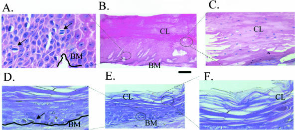
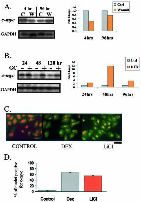
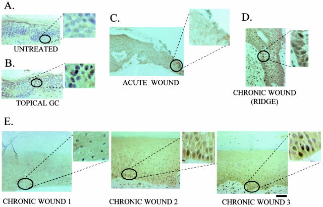
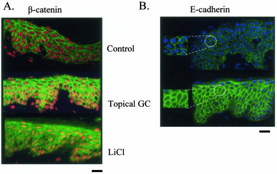
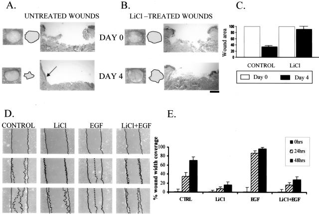
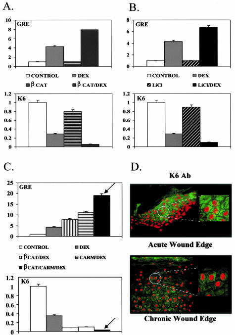
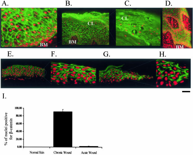
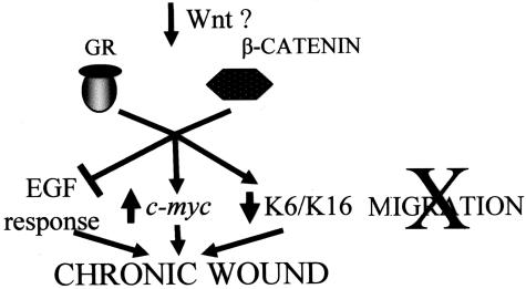
Lack of understanding of the molecular mechanisms and pathogenesis of impaired healing in chronic ulcers is a serious health issue that contributes to excessive limb amputations and mortality. Here we show that beta-catenin and its downstream targets in keratinocytes, c-myc, and keratins K6 and K16, play important roles in the development of chronic wounds. In contrast to normal epidermis, we observed a significant nuclear presence of beta-catenin and elevated c-myc expression at the nonhealing wound edge of chronic ulcers from 10 patients. In vitro studies indicated that stabilization of nuclear beta-catenin inhibited wound healing and keratinocyte migration by blocking epidermal growth factor response, inducing c-myc and repressing the K6/K16 keratins (cytoskeletal components important for migration). The molecular mechanism of K6/K16 repression involved beta-catenin and arginine methyltransferase (CARM-1) acting as co-repressors of glucocorticoid receptor monomers. We conclude that activation of the beta-catenin/c-myc pathway(s) contributes to impaired healing by inhibiting keratinocyte migration and altering their differentiation. The presence of activated beta-catenin and c-myc in the epidermis of chronic wounds may serve as a molecular marker of impaired healing and may provide future targets for therapeutic intervention.


Figures

Figure 1
Figure 1
Histology of chronic wounds is consistent with activation of c-myc. A to C: Histology of chronic ulcers. A: Higher magnification shows mitotically active cells found in suprabasal layers (arrows) indicating aberrant proliferation. B: Low magnification shows thickened, hyperproliferative, hyper- and parakeratotic epidermis. C: High magnification shows parakeratosis (nuclei present in cornified layer) indicating inappropriate differentiation. D to F: Histology of normal skin. D: Mitosis only in basal layer of epidermis; E: low magnification; F: cornified layer, high magnification. BM, basal membrane; CL, cornified layer.
Figure 2
Figure 2
c-myc is differentially regulated by wound healing and its inhibitor, GC. Northern blots with mRNA isolated from acute wounds 4 and 96 hours after wounding (A) and topical GC treatment of human skin (B). c-myc was repressed in early wound healing, while induced by GC. Bar graphs show quantification of the Northern blots by densitometry. C: Immunofluorescence of primary human keratinocytes incubated with GC and LiCl stained with c-myc-specific antibody. To better visualize its nuclear presence we counterstained the nuclei. c-myc is visualized by green fluorescein isothiocyanate and its nuclear presence changes their color from red (see control) to orange/yellow (see treated cells). Both GC (middle) and LiCl, ie, stabilized β-catenin (right) induce c-myc as evident by positive nuclear staining. D: Graph represents average ± SD of percent of nuclei positive for c-myc for several independent experiments.
Figure 3
Figure 3
c-myc as a marker of inhibition of wound healing in chronic ulcers in vivo. Immunohistochemistry of skin samples stained with c-myc-specific antibody. A: Normal human skin shows absence of c-myc. B: Skin treated with topical GC shows induction of c-myc. C: Epidermis at the edge of an acute wound shows absence of c-myc, thus confirming that c-myc is not activated in acute wound healing. D and E: c-myc is induced in chronic ulcers in vivo. D: Most prominent induction of c-myc in the basal layer of epidermis; E: c-myc is activated throughout the epidermis at the nonhealing edge of a chronic ulcers. Insets show enlarged images of nuclear staining.
Figure 4
Figure 4
Topical GC activates β-catenin pathway in the epidermis of human skin. A: Immunofluorescence of normal human skin treated either with topical GC or LiCl (positive control) reveals nuclear β-catenin (visualized by orange/yellow nuclei) in treated skin whereas it is on the membrane in untreated skin (red nuclei indicate absence of signal). B: Immunofluorescence shows that E-cadherin remains membrane-associated in untreated skin whereas it becomes internalized (cytoplasmic) in GC-treated skin. Insets show enlarged images.
Figure 5
Figure 5
Activation of β-catenin inhibits wound healing and keratinocyte migration. LiCl (ie, nuclear β-catenin) causes delayed wound healing in human skin organ culture wounds. Both gross pictures of untreated (A) and LiCl-treated wounds (B) as well as their histology are shown. Arrow points at epithelial tongue indicating active healing in untreated wounds whereas it is absent in the LiCl-treated wounds. Filled circles indicate the wound surfaces. C: Quantification of the wound size by planimetry shows 70% healing rate of untreated wounds and only 12% healing rate for LiCl-treated wounds. D: LiCl inhibits migration of primary human keratinocytes in wound scratch assay when compared with untreated cells. Inhibition is prominent even in first 24 hours and was further sustained through 48 hours. EGF (positive control) stimulated migration and wound was completely closed after 48 hours. Importantly, this activation of endogenous β-catenin completely blocked EGF-stimulated migration. Full lines indicate initial wound area; dotted lines demarcate migrating front of cells. E: Histograms indicate the average coverage of scratch wounds widths in percent relative to baseline wound width at the day 0 and 24 and 48 hours after LiCl, EGF, and LiCl/EGF treatments.
Figure 6
Figure 6
Molecular mechanism of inhibition of keratinocyte migration by β-catenin involves GR and CARM-1 and cytoskeletal component K6 keratin. A: Graph represents quantitative CAT assay after co-transfection of human keratinocytes with K6-CAT promoter showing that β-catenin enhances repression of K6 by the GC, dexamethasone (DEX), thus acting as a co-repressor of GR. B: Similarly, LiCl treatment (ie, endogenously activated β-catenin) also enhances repression of K6 by DEX. C: The β-catenin-mediated co-repression is further enhanced by arginine methyltransferase CARM-1 (arrow), indicating that GR, β-catenin, and CARM-1 act as a repressor complex that suppresses K6. D: Sections of chronic wounds stained with K6-specific antibody (bottom) revealed marked repression of K6 levels when compared with acute wound (top).
Figure 7
Figure 7
β-Catenin is activated in chronic wounds in vivo. Immunofluorescence with β-catenin-specific antibody. Prominent nuclearization of β-catenin is found in epidermis at the nonhealing edge of a chronic ulcer. Higher magnification shows nuclear presence of β-catenin in epidermis (A) and in parakeratotic cornified layer (C). B: Low magnification shows nuclear β-catenin signal throughout epidermis. D: The nuclearization signal is strongest toward basal layers, around granulation tissue of the actual edge. E and F: Control: normal skin. Normal epidermis of human skin shows β-catenin localized only to the membrane and not in the nucleus: low magnification (E) and enlarged (F). G and H: Control: acute wound. Epidermis at the edge of the acute wound shows β-catenin localized only at the membrane and not in the nucleus. G: Low magnification and H: enlarged. I: Histogram represents average ± SD of percent of nuclei positive for β-catenin from two different ulcers and several different sections.
Figure 8
Figure 8
Cartoon summarizes our findings suggesting the molecular mechanism through which β-catenin and c-myc may participate in development of chronic wounds.

Similar articles

Cited by

References

    1. Jamora C, DasGupta R, Kocieniewski P, Fuchs E. Links between signal transduction, transcription and adhesion in epithelial bud development. Nature. 2003;422:317–322. - PMC - PubMed
    1. Kirfel J, Magin TM, Reichelt J. Keratins: a structural scaffold with emerging functions. Cell Mol Life Sci. 2003;60:56–71. - PMC - PubMed
    1. Tomic-Canic M, Magnus SA, Oscar MA. Epidermal repair and the chronic wound. Rovee DT, Maibach HI, editors. Boca Raton: CRC Press LLC,; The epidermis in wound healing. 2004:pp 25–57.
    1. Morasso MI, Tomic-Canic M. Epidermal stem cells: the cradle of epidermal determination, differentiation and wound healing. Biol Cell. 2005;97:173–183. - PMC - PubMed
    1. Jeffcoate WJ, Harding KG. Diabetic foot ulcers. Lancet. 2003;361:1545–1551. - PubMed

Publication types

MeSH terms