

Expression profiles and functional analyses of Wnt-related genes in human joint disorders
- PMID: 15972956
- PMCID: PMC1603448
- DOI: 10.1016/S0002-9440(10)62957-4
Expression profiles and functional analyses of Wnt-related genes in human joint disorders
Abstract
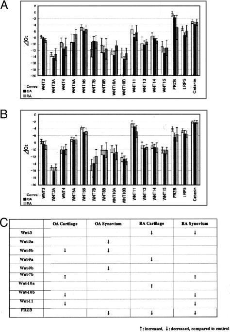
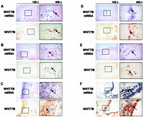
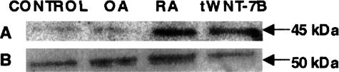
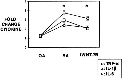
Rheumatoid arthritis (RA) and osteoarthritis (OA) are joint disorders that cause major public health problems. Previous studies of the etiology of RA and OA have implicated Wnt genes, although the exact nature of their involvement remains unclear. To further clarify the relationship between RA, OA, and the Wnt gene family, gene expression analyses were performed on articular cartilage, bone, and synovial tissues in knee joints taken from RA, OA, and nor-mal/control patients. Cytokine assays were also performed in cells transfected with Wnt-7b, a member of the gene family most closely linked to RA and OA. Of the human Wnt genes, real-time PCR analysis revealed significant up-regulation of Wnt-7b in OA cartilage and RA synovium. In situ hybridization and immunohistochemistry also revealed that Wnt-7b was present in articular cartilage, bone, and synovium of RA samples and in osteophytes, articular cartilage, bone marrow, and synovium of OA samples. The levels of the cytokines tumor necrosis factor-alpha, interleukin-1beta, and interleukin-6 were significantly increased in RA synovium and Wnt-7b-transfected normal synovial cells when compared with normal samples. These results point to the potential involvement of Wnt signaling in the pathobiology of both RA and OA.
Figures
Comment in
-
Wnt signaling and orthopedic diseases.Am J Pathol. 2005 Jul;167(1):1-3. doi: 10.1016/S0002-9440(10)62947-1. Am J Pathol. 2005. PMID: 15972946 Free PMC article. Review. No abstract available.
Similar articles
-
Differential expression of WNTs and FRPs in the synovium of rheumatoid arthritis and osteoarthritis.Biochem Biophys Res Commun. 2006 Jul 14;345(4):1615-20. doi: 10.1016/j.bbrc.2006.05.075. Epub 2006 May 22. Biochem Biophys Res Commun. 2006. PMID: 16735027
-
Responses to the proinflammatory cytokines interleukin-1 and tumor necrosis factor alpha in cells derived from rheumatoid synovium and other joint tissues involve nuclear factor kappaB-mediated induction of the Ets transcription factor ESE-1.Arthritis Rheum. 2003 May;48(5):1249-60. doi: 10.1002/art.10942. Arthritis Rheum. 2003. PMID: 12746898
-
Comparison of cathepsins K and S expression within the rheumatoid and osteoarthritic synovium.Arthritis Rheum. 2002 Mar;46(3):663-74. doi: 10.1002/art.10114. Arthritis Rheum. 2002. PMID: 11920402
-
Novel insight into the role of α-actinin-1 in rheumatoid arthritis.Discov Med. 2014 Feb;17(92):75-80. Discov Med. 2014. PMID: 24534470 Review.
-
Activated synovial macrophages as targets for osteoarthritis drug therapy.Curr Drug Targets. 2010 May;11(5):576-85. doi: 10.2174/138945010791011965. Curr Drug Targets. 2010. PMID: 20199392 Review.
Cited by
-
Targeting Wnt pathways in disease.Cold Spring Harb Perspect Biol. 2012 Nov 1;4(11):a008086. doi: 10.1101/cshperspect.a008086. Cold Spring Harb Perspect Biol. 2012. PMID: 23001988 Free PMC article. Review.
-
Disease-modifying therapeutic strategies in osteoarthritis: current status and future directions.Exp Mol Med. 2021 Nov;53(11):1689-1696. doi: 10.1038/s12276-021-00710-y. Epub 2021 Nov 30. Exp Mol Med. 2021. PMID: 34848838 Free PMC article. Review.
-
Relationship between joint shape and the development of osteoarthritis.Curr Opin Rheumatol. 2010 Sep;22(5):538-43. doi: 10.1097/BOR.0b013e32833d20ae. Curr Opin Rheumatol. 2010. PMID: 20644480 Free PMC article. Review.
-
FGF23 Regulates Wnt/β-Catenin Signaling-Mediated Osteoarthritis in Mice Overexpressing High-Molecular-Weight FGF2.Endocrinology. 2018 Jun 1;159(6):2386-2396. doi: 10.1210/en.2018-00184. Endocrinology. 2018. PMID: 29718273 Free PMC article.
-
Dendritic Cell-Specific Deletion of β-Catenin Results in Fewer Regulatory T-Cells without Exacerbating Autoimmune Collagen-Induced Arthritis.PLoS One. 2015 Nov 20;10(11):e0142972. doi: 10.1371/journal.pone.0142972. eCollection 2015. PLoS One. 2015. PMID: 26587585 Free PMC article.
References
-
- Haugeberg G, Orstavik RE, Kvein TK. Effects of rheumatoid arthritis on bone. Curr Opin Rheumatol. 2003;15:469–475. - PubMed
-
- Moskowitz RW, Howell DS, Altman RD, Buckwalter JA, Goldberg VM. Osteoarthritis, ed 3. Philadelphia: W.B. Saunders,; Diagnosis and Medical/Surgical Management, 2003
-
- Hashimoto S, Creghton-Achermann, Takahashi K, Amiel D, Coutts RD. Development and regulation of osteophyte formation during experimental osteoarthritis. Osteoarthritis Cartilage. 2002;10:180–187. - PubMed
-
- National Center for Health Statistics, Centers for Disease Control and Prevention, US Department of Health and Human Services Hyattsville, MD: Division of Data Services, National Center for Health Statistics, US Centers for Disease Control and Prevention,; National Health and Nutrition Examination Survey, III 1988–1994. 1996
-
- Grotewold L, Ruther U. Bmp, Fgf and Wnt signalling in programmed cell death and chondrogenesis during vertebrate limb development: the role of Dickkopf-1. Int J Dev Biol. 2002;46:943–947. - PubMed
Publication types
MeSH terms
Substances
LinkOut - more resources
Full Text Sources
Medical
Research Materials
