. 2005 Jul;167(1):151-9.
doi: 10.1016/S0002-9440(10)62962-8.

Perturbed neurogenesis in the adult hippocampus associated with presenilin-1 A246E mutation

Affiliations

Perturbed neurogenesis in the adult hippocampus associated with presenilin-1 A246E mutation

Nathalie L Chevallier et al. Am J Pathol. 2005 Jul.

Abstract

In addition to its well-established role in gamma-secretase cleavage, presenilin (PS) also plays a role in regulating the stability of cytosolic beta-catenin, a protein involved in Wnt signaling. Several familial Alzheimer's disease-associated PS1 mutations have been shown to increase the stability of the signaling pool of beta-catenin, correlating with enhanced cell proliferation. Accordingly, we hypothesized that in the setting of PS1 mutations, abnormal activation of Wnt/beta-catenin signaling leads to increased cell division. We tested this hypothesis by examining whether there is evidence of increased neurogenesis in the hippocampus of adult transgenic mice that overexpress the PS1 A246E mutation. In PS1/PS2-deficient fibroblasts, expression of PS1 A246E Familial AD mutation failed to restore the rapid turnover of beta-catenin compared with wild-type PS1. We then examined whether the same mutation enhanced neurogenesis in vivo in adult hippocampus of PS1-deficient mice when restored by wild-type human PS1 (PS1(-/-)WT) or A246E PS1 mutation (PS1(-/-)AE). The PS1 A246E mutation stimulated the proliferation of progenitor cells in the dentate gyrus of adult mice, as assessed by 5-bromo-2-deoxyuridine incorporation, but did not influence their survival or differentiation. These observations suggest that the PS1 A246E mutation influences cell growth putatively via abnormal beta-catenin signaling in vivo.


Figures

Figure 1
Figure 1
Defective turnover of β-catenin in cells genetically deficient in mouse presenilins and rescued by hPS1-bearing FAD-linked mutations. A: Confluent cultures of PS−/− cells and PS−/− cells reconstituted with either wild-type PS1 or FAD mutations were treated with 25 μmol/L cycloheximide (CHX) for the indicated time periods and immunoblotted for P-45 β-catenin. Immunoblot exposures were adjusted to show similar signal at time 0. B: The graph represents the relative turnover in P-45 β-catenin from three independent experiments. Error bars represent SEM.
Figure 2
Figure 2
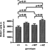
Increased cell proliferation in the adult dentate gyrus of mice genetically deficient in mouse PS1 and transgenic for PS1A246E. The numbers of BrdU-positive cells are similar in PS1+/+ and PS1−/−WT animals but significantly lower than PS1−/−AE and PS1+/− animals. Results shown are means of total numbers of BrdU-positive cells (±SEM) for each genotype studied (n = 5 to 7 mice per group). Statistical significance was evaluated using analysis of variance followed by Newman-Keuls post hoc multiple comparison test.
Figure 3
Figure 3
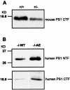
Expression of PS1 transgenic protein in PS1 wild-type and PS1 A246E mice. Immunoblots of brain homogenates reacted with the 490 antibody (human and mouse PS1 C-terminal fragment) and PSN2 antibody (human-specific PS1 N-terminal fragment). A: The level of endogenous mouse PS1 C-terminal fragment is reduced in PS1+/− brain compared with PS1+/+. B: The expression level of both N- and C-terminal PS1 fragments is approximately 40% higher in PS1−/−A246E mice compared with PS1 wild-type mice (n = 3 mice per genotype).
Figure 4
Figure 4
Expression of human PS1 transgenic protein in progenitor cells. Immunostaining of human PS1 using a human-specific rat anti-PS1 monoclonal antibody 24-4B5 was performed the day after BrdU injections. A: Specific human PS1 immunostaining is seen diffusely in the dentate gyrus of a PS1−/−AE mouse (tg, transgenic; top panel) but not in a control PS1+/+ nontransgenic animal (non-tg; bottom panel). gcl, granular cell layer of the dentate guys. Scale bar = 100 μm. B: PS1 (top panel. red) is expressed in the subgranular zone (sgz) where dividing progenitor cells are located (bottom panel, green for BrdU). Arrowheads, BrdU-positive cells. Note that due to the thickness of the section (40 μm), only a few BrdU cells are in the plane of focus. Pictures in B are a close-up view of the rectangle highlighted in A. Scale bar = 25 μm. C: In higher magnification, representative confocal laser scanning microscopy images showing colocalization of a neuron immunolabeled for PS1 and BrdU. The bottom panel [BdU] represents an overexposure of the BrdU photomicrograph shown in the second panel to demonstrate location of BrdU-negative cells (asterisks). Systematic assessment of double-labeled images by confocal laser scanning microscopy study confirmed that all of the BrdU-positive cells were immunopositive for human PS1. Scale bar = 10 μm.
Figure 5
Figure 5
Survival of BrdU cells 4 weeks after injections is comparable between the different experimental groups. The total number of BrdU-positive cells (means of total ± SEM) were similar among all four genotypes (P > 0.05, n = 5 to 10 animals per group).
Figure 6
Figure 6
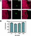
Neuronal differentiation of progenitor cells is unchanged by PS1 mutation. Double immunofluorescent labeling of BrdU and NeuN was performed on brain sections 4 weeks after BrdU injections to confirm neuronal differentiation. A: NeuN-positive cells (red) decorate the granule cell layer of the dentate gyrus. Newly differentiated neurons are double-labeled for BrdU (green) and NeuN (red) (top panels). Bottom panels: An example of a BrdU-positive cell that did not show NeuN immunostaining and presumably failed to differentiate in a neuron. Scale bar = 10 μm. B: Neuronal differentiation of progenitor cells was similar between the four experimental groups. The percent BrdU cells that are double-labeled for NeuN was scored from the animals illustrated in A (P > 0.05, n = 4 to 6 per group). No statistical differences were found between any of the studied genotypes.

References

    1. St George-Hyslop PH. Molecular genetics of Alzheimer’s disease. Biol Psychiatry. 2000;47:183–199. - PubMed
    1. Duff K, Eckman C, Zehr C, Yu X, Prada CM, Perez-tur J, Hutton M, Buee L, Harigaya Y, Yager D, Morgan D, Gordon MN, Holcomb L, Refolo L, Zenk B, Hardy J, Younkin S. Increased amyloid-beta42(43) in brains of mice expressing mutant presenilin 1. Nature. 1996;383:710–713. - PubMed
    1. Scheuner D, Eckman C, Jensen M, Song X, Citron M, Suzuki N, Bird TD, Hardy J, Hutton M, Kukull W, Larson E, Levy-Lahad E, Viitanen M, Peskind E, Poorkaj P, Schellenberg G, Tanzi R, Wasco W, Lannfelt L, Selkoe D, Younkin S. Secreted amyloid beta-protein similar to that in the senile plaques of Alzheimer’s disease is increased in vivo by the presenilin 1 and 2 and APP mutations linked to familial Alzheimer’s disease. Nat Med. 1996;2:864–870. - PubMed
    1. Wen PH, Friedrich VL, Jr, Shioi J, Robakis NK, Elder GA. Presenilin-1 is expressed in neural progenitor cells in the hippocampus of adult mice. Neurosci Lett. 2002;318:53–56. - PubMed
    1. Busciglio J, Hartmann H, Lorenzo A, Wong C, Baumann K, Sommer B, Staufenbiel M, Yankner BA. Neuronal localization of presenilin-1 and association with amyloid plaques and neurofibrillary tangles in Alzheimer’s disease. J Neurosci. 1997;17:5101–5107. - PMC - PubMed

Publication types

MeSH terms