. 2005 Aug;3(8):e242.
doi: 10.1371/journal.pbio.0030242. Epub 2005 Jun 28.

The Ews-ERG fusion protein can initiate neoplasia from lineage-committed haematopoietic cells

Affiliations

The Ews-ERG fusion protein can initiate neoplasia from lineage-committed haematopoietic cells

Rosalind Codrington et al. PLoS Biol. 2005 Aug.

Abstract

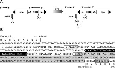
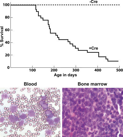
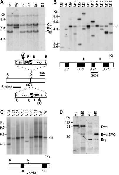
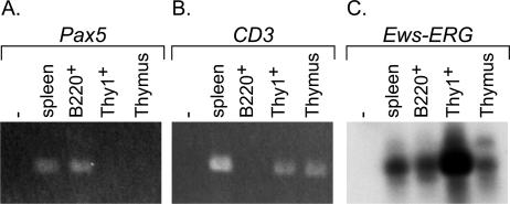
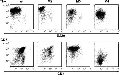
The EWS-ERG fusion protein is found in human sarcomas with the chromosomal translocation t(21;22)(q22;q12), where the translocation is considered to be an initiating event in sarcoma formation within uncommitted mesenchymal cells, probably long-lived progenitors capable of self renewal. The fusion protein may not therefore have an oncogenic capability beyond these progenitors. To assess whether EWS-ERG can be a tumour initiator in cells other than mesenchymal cells, we have analysed Ews-ERG fusion protein function in a cellular environment not typical of that found in human cancers, namely, committed lymphoid cells. We have used Ews-ERG invertor mice having an inverted ERG cDNA cassette flanked by loxP sites knocked in the Ews intron 8, crossed with mice expressing Cre recombinase under the control of the Rag1 gene to give conditional, lymphoid-specific expression of the fusion protein. Clonal T cell neoplasias arose in these mice. This conditional Ews gene fusion model of tumourigenesis shows that Ews-ERG can cause haematopoietic tumours and the precursor cells are committed cells. Thus, Ews-ERG can function in cells that do not have to be pluripotent progenitors or mesenchymal cells.


Figures

Figure 1
Figure 1. Strategy for Generating Ews-ERG Invertor Gene by Homologous Recombination
(A) The method for making invertor mice is described in detail elsewhere [35]. In summary, an invertor cassette comprising a short intronic region with an acceptor splice site, human ERG cDNA sequence (shown in Figure S1), a polyA site, and the MC1neopA gene all flanked by loxP sites (depicted by black triangles) was knocked in, using homologous recombination, into Ews intron 8. The transcription orientation of the targeted ERG cDNA invertor cassette was opposite from that of the endogenous Ews gene after initial homologous recombination. Germ line carrier mice of this targeted allele were crossed with Cre-expressing mouse strains [36], and, as indicated in the right hand side of the diagram, the invertor cassette is turned around to create a transcription orientation identical with that of Ews. Thus, after transcription, a pre-mRNA is made with the donor splice site of Ews exon 7 adjacent to the acceptor site of the invertor cassette, allowing post-transcriptional fusion of Ews with ERG in an analogous format to that found in human sarcomas with t(21;22). (B) Genomic sequence adjacent to the Ews exon 7 and the ERG invertor cassette (the derived amino acid sequence is shown in the single letter code) obtained from DNA of ES cells and of thymus after Cre-mediated inversion. The Ews intron 7/8 donor splice site is indicated by an arrow. The boxed sequence ( TCTAG/ CGAT) corresponds to the ligation of filled-in XbaI (Ews genomic) and ClaI (ERG invertor cassette) sites (for detailed description see [35]). The boxed XbaI site ( TCTAGA) corresponds to the position of cloning of the mouse Af4 intronic sequence (shaded) in the ERG invertor cassette. On the 3′ side of the fused XbaI-ClaI sites, there is a loxP site originating from the ERG invertor cassette (see Figure S1) followed by the Af4 intron 4 (shaded) upstream of the ERG cDNA sequence. The Af4 acceptor splice site is indicated by an arrow, and is followed by the ERG cDNA sequence. A NotI site used for cloning the amplified ERG sequence is boxed (see also Figure S1) (note this sequence is non-contiguous and the dots represent a gap; the full sequence appears in [35]).
Figure 2
Figure 2. Incidence of Haematological Malignancy and Characteristics of Ews-ERG Invertor Mice
Cohorts of 29 Ews-ERG; Rag1-Cre mice and 20 Ews-ERG control mice were monitored over a period of approximately 17 mo. Mice were culled and a post-mortem conducted when signs of ill health were observed. Leukaemia/lymphoma was established by various criteria (see Materials and Methods). Top panel shows the survival curve of Ews-ERG; Rag1-Cre (+Cre) or Ews-ERG (−Cre) mice as a function of time (days). Bottom panel shows the histology of blood and bone marrow from leukaemic mice (M21 and M20, respectively). Blood smears were stained with May-Grünwald-Giemsa stain and photographed at 400× magnification, whilst bone marrow was photographed at 1,000×.
Figure 3
Figure 3. Clonality of T Cell Neoplasias inEws-ERG Invertor Mice
Genomic DNA was prepared from various tissues of invertor mice with thymomas. Genomic analysis was carried out using filter hybridisation to assess the presence of the inverted ERG cassette in the tumour cells (A), and to assess whether the lymphocytes involved in the tumours were clonal T (B) or B cells (C). (A) Inversion hybridisation autoradiograph. DNA was prepared from the thymoma and other tissues of M18, cleaved with EcoRI and hybridised to the 5′ Ews probe (which detects a 5-kb targeted ERG cassette fragment or a 6.5-kb Cre-inverted fragment). If the ERG cassette is inverted by Cre activity, the size of the EcoRI fragment increases from the initial targeted gene size, as indicated in the maps below the figure. The data shown are for DNA extracted from spleen (spl), thymus (thy), liver (liv), kidney (kid) or tail (ES cell DNA is used as a control). The Cre-mediated inverted band (∼6.5 kb) is evident in thymus DNA (thymoma). The summary of the data from the cohort of invertor mice is in Table 2. Note that despite extensive infiltration of spleen detected by histology, we cannot see the inverted band by Southern blot; this presumably reflects regional clustering of neoplastic cells in spleen. Restriction fragment sizes are represented as germ line (GL), inverted allele of ERG cassette (inv) and initial targeted Ews allele (Tgt). The organisation of the targeted Ews allele is indicated underneath. The hybridisation autoradiograph shows the location of Ews exon 7 and the initial targeted orientation (bottom) or inverted orientation of ERG invertor cassette after Cre-mediated recombination (top). a, acceptor splice site. (B) Autoradiograph showing rearrangement of T cell receptor β locus. A T cell receptor Jβ2 probe [40] (diagrammatically shown below the autoradiograph) detects a 5-kb germ line HindIII Jβ2 band whilst V-D-Jβ2 joins in T cells result in new HindIII-sized bands depending on the nature of the rearrangement. Each of 12 thymoma DNAs that were compared showed one or two Jβ2 alleles rearranged, signifying that these tumours were clonal T cells. In some thymoma samples, there is almost complete absence of the germ line band, indicating that the thymuses of these mice are solely comprised of malignant clonal T cells. DNA sequence analysis of the V-D-J junctions of mice M2, M5, M13, and M18 showed functional V-D-J joins (see Table 2). (C) Autoradiograph showing rearrangement status of immunoglobulin heavy-chain genes. A Cμ intron probe [41] (diagrammatically shown below the autoradiograph) was used to hybridise a set of thymoma DNAs for the presence of Igh rearranged bands. Only two samples showed rearrangements. (D) Detection of Ews-Erg fusion protein in thymoma cells. Single cell suspensions were made of T cells from a normal thymus (wt) or from the thymoma of M6, protein fractionated on 4%–20% acrylamide gel, and transferred to nylon membranes. Specific proteins were detected with anti-Ews or anti-ERG antibody.
Figure 4
Figure 4. B and T Cells Express the Ews-ERG Fusion RNA
A 96-d-old mouse with both Ews-ERG and Rag1-Cre alleles was used as a source of spleen and thymus cells. Single cell suspensions of spleen cells were labelled with anti-B220 or with anti-Thy1.2 and were purified using a MoFlo preparative flow cytometer. Estimated purities were achieved of greater than 95%. cDNA was prepared from RNA extracted from sorted cells or from aliquots of unsorted populations and RT-PCR (approximately 3,400 B220+ or 6,400 Thy1.2+ cell equivalents per PCR reaction) carried out with specific Pax5 (A), CD3 (B) or Ews-ERG (C) primers. PCR reaction products were fractionated on 1% agarose gels and either stained with ethidium bromide and photographed (A and B) or gel blotted and hybridised with an Ews-ERG probe (C)
Figure 5
Figure 5. Analysis of Cell Surface Antigens of Thymomas from Ews-ERG Invertor Mice Using Flow Cytometry
Thymus tissue was resected from mice with thymoma and FACS analysis performed to determine T cell phenotype (summarised in Table 1). The data in the figure show representative flow diagrams of three tumour-bearing mice (M2, M3, and M4) compared with a wild-type C57BL/6 control (wt), using anti-Thy1 (y-axis) plus anti-B220 (x-axis) antibodies or using anti-CD8 (y-axis) plus anti-CD4 (x-axis) antibodies. The three thymomas show a range of CD4 and CD8 co-expression phenotypes characterizing the thymomas generated in the Ews-ERG; Rag1-Cre invertor mice..

Similar articles

Cited by

References

    1. Rabbitts TH. Chromosomal translocations in human cancer. Nature. 1994;372:143–149. - PubMed
    1. Look AT. Oncogenic transcription factors in the human acute leukemias. Science. 1997;278:1059–1065. - PubMed
    1. Mitelman F, Johansson B, Mertens F, editors. Mitelman Database of Chromosome Aberrations in Cancer [database] Bethesda (Maryland): National Cancer Institute; 2000. Available: http://cgap.nci.nih.gov/Chromosomes/Mitelman. Accessed 19 May 2005.
    1. Huntly BJ, Gilliland DG. Leukaemia stem cells and the evolution of cancer-stem-cell research. Nat Rev Cancer. 2005;5:311–321. - PubMed
    1. Gale RP, Grosveld G, Canaani E, Goldman JM. Chronic myelogenous leukemia: Biology and therapy. Leukemia. 1993;7:653–658. - PubMed

Publication types

MeSH terms

LinkOut - more resources