. 2005 Nov 1;391(Pt 3):513-25.
doi: 10.1042/BJ20050491.

Ubiquilin regulates presenilin endoproteolysis and modulates gamma-secretase components, Pen-2 and nicastrin

Affiliations

Ubiquilin regulates presenilin endoproteolysis and modulates gamma-secretase components, Pen-2 and nicastrin

Leann K Massey et al. Biochem J. .

Abstract

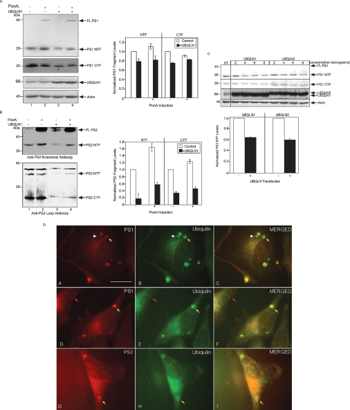
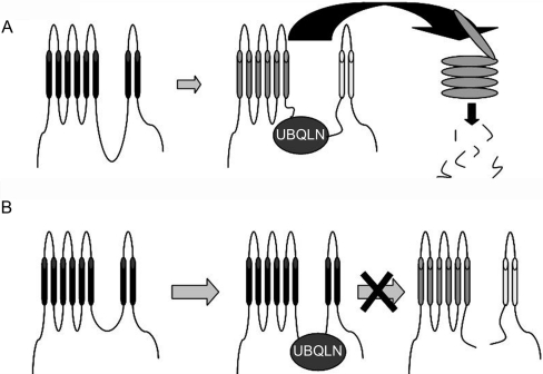
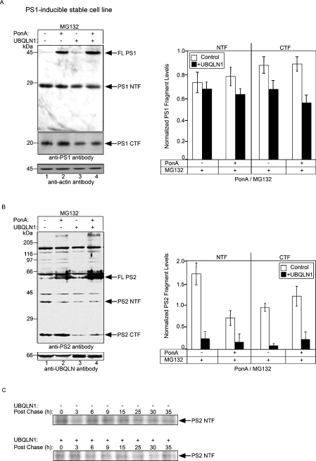
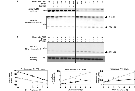
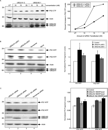
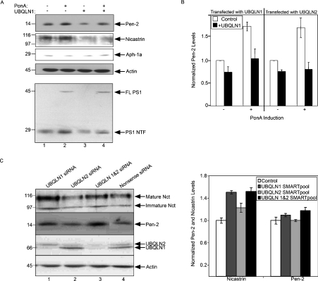
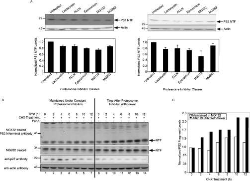
Mutations in presenilin proteins (PS1 and PS2) lead to early-onset Alzheimer's disease. PS proteins are endoproteolytically cleaved into two main fragments: the NTF (PS N-terminal fragment) and the CTF (PS C-terminal fragment). The two fragments are believed to constitute the core catalytic enzyme activity called gamma-secretase, which is responsible for cleaving beta-amyloid precursor protein to release Abeta. Thus, studying factors that modulate PS fragment levels could provide important information about gamma-secretase. Previously, we demonstrated that the protein, ubiquilin-1, interacts both in vivo and in vitro with PS and that overexpression of ubiquilin-1 or -2 leads to increased accumulation of full-length PS proteins. Using wild-type HEK-293 cells (human embryonic kidney 293 cells) and PS-inducible cells, we now show that overexpression of either ubiquilin-1 or -2 decreases the PS NTF and CTF levels. Conversely, siRNA (small interfering RNA)-mediated knockdown of ubiquilin-1 and -2 proteins increased the PS NTF and CTF levels. We considered that ubiquilin might alter PS fragment accumulation by acting as a shuttle factor escorting PS fragments to the proteasome for degradation. However, through proteasome inhibition studies, we show that this does not occur. Instead, our results suggest that ubiquilin regulates PS fragment production. We also examined whether other components of the gamma-secretase complex are affected by ubiquilin expression. Interestingly, overexpression of ubiquilin resulted in a decrease in Pen-2 and nicastrin levels, two essential components of the gamma-secretase complex. In contrast, knockdown of ubiquilin-1 and -2 protein expression by RNAi (RNA interference) increased Pen-2 and nicastrin levels. Finally, we show that inhibition of the proteasome results in decreased PS fragment production and that reversal of proteasome inhibition restores PS fragment production, suggesting that the proteasome may be involved in PS endoproteolysis. These studies implicate ubiquilin as an important factor in regulating PS biogenesis and metabolism.


Figures

Figure 1
Figure 1. Differential modulation of FL and PS protein fragments by ubiquilin-1 in HEK-293 PS-inducible cell lines
(A) PS1 NTF and CTF levels were analysed in PS1 cell lines that were either left uninduced (lanes 1 and 3) or induced with PonA (lanes 2 and 4) and either left untransfected (lanes 1 and 2) or transfected with a ubiquilin-1 expression construct (UBQLN1; lanes 3 and 4). Equivalent amounts of protein lysates were separated by SDS/PAGE (10%, w/v, polyacrylamide) and immunoblotted with anti-PS1 NTF antibody and anti-PS1 loop antibody. The filter was then reprobed with anti-ubiquilin antibody to ensure overexpression of ubiquilin and with an anti-actin antibody to ensure equal protein loading. Note that overexpression of ubiquilin-1 decreases PS1 NTF and CTF levels (lanes 3 and 4). Densitometric analysis was used to quantify the extent of PS fragment reduction. Each experiment was repeated more than three times. (B) Same experiment as described in (A) except that PS2-inducible cells were used to analyse PS2 NTF and CTF levels. Like PS1 fragments, overexpression of ubiquilin-1 decreased PS2 fragment levels. The nature of the 40 kDa band is unknown; however, it appears to be a PS2-related fragment because its levels also decreased upon ubiquilin overexpression. (C) Endogenous PS fragment levels were analysed in HEK-293 cells that were transfected with increasing amounts of ubiquilin-1 or -2 expression plasmids. Equivalent amounts of protein lysates were separated by SDS/PAGE (10% polyacrylamide) and immunoblotted with an anti-PS1 NTF antibody. The filter was then reprobed with anti-ubiquilin antibody to ensure overexpression of ubiquilin and with an anti-actin antibody to ensure equal protein loading. Note that FL PS1 is not visible in these cells. Similar to PS2, overexpression of ubiquilin decreased PS1 NTF levels and the extent of reduction was quantified again by densitometric analysis. The graph shows the quantification of PS1 fragments in cells that were transfected with 8 μg of ubiquilin expression plasmid compared with untransfected (UT) cells. (D) Endogenous ubiquilin and PS proteins co-localize in vesicular-like structures in HeLa cells. HeLa cells were double stained with rat monoclonal anti-PS1 antibody (A and D) and with a rabbit anti-ubiquilin N-terminal antibody (B and E) or with a goat anti-PS2 antibody (G) and a mouse anti-ubiquilin antibody (H). A merged image of the two primary images is shown to the right of each set. Arrows indicate puncta of unknown origin in which PS and ubiquilin staining was co-localized. Scale bar, 5 μm (D, A–H).
Figure 2
Figure 2. Potential mechanisms by which ubiquilin decreases PS fragment levels
(A) FL PS is cleaved into its fragments. The PS fragments then interact with ubiquilin (UBQLN), then escorts the fragments to the proteasome for rapid degradation. (B) Ubiquilin interacts with FL PS, blocking access of the presenilinase responsible for endoproteolysis.
Figure 3
Figure 3. Overexpression of ubiquilin does not facilitate rapid degradation of PS fragments
(A) PS1 cell lines that were either left uninduced (lanes 1 and 3) or induced with PonA (lanes 2 and 4) and either left untransfected (lanes 1 and 2) or transfected with ubiquilin-1 expression plasmids (lanes 3 and 4) were treated for 7 h with MG132 after which protein lysates were collected and analysed by immunoblotting. Even after MG132 treatment, PS1 NTF and CTF levels remain reduced in transfected cells (compare lanes 3 and 1 or lanes 4 and 2), indicating that ubiquilin does not decrease PS fragment levels by increasing the rate of proteasome-dependent degradation of the fragments. For clarification, if overexpression of ubiquilin was to increase PS fragment degradation via the proteasome, then, on proteasome inhibition, PS fragment turnover would be blocked and PS fragment levels would be similar, or increase, relative to that of untransfected cells. However, this was not the case, suggesting that ubiquilin does not increase PS fragment turnover. Densitometric analysis was used to quantify the extent of reduction. Each experiment was repeated more than three times and similar trends were observed. (B) Same experiment as described in (A) except that PS2-inducible cell lines were used to examine PS2 NTF and CTF levels. (C) Ubiquilin-1 overexpression does not alter the turnover rate of PS2 NTF in PS2-inducible cells. Classical [35S]methionine pulse–chase study of immunoprecipitated PS2 NTF in cells in which ubiquilin was overexpressed compared with untransfected cells. The rate of PS2 NTF turnover was calculated to be the same irrespective of ubiquilin-1 overexpression.
Figure 4
Figure 4. Overexpression of ubiquilin reduces PS fragment production
(A) A time-course experiment using ponA-induced PS2 cell line cultures that were transfected with or without ubiquilin-1 (UBQLN) expression plasmids and treated with 100 μM cycloheximide (CHX) for 0–6 h. Equal amounts of protein lysate were separated by SDS/PAGE (8.5% polyacrylamide) and subsequently immunoblotted with anti-ubiquilin antibody. These blots illustrated that the transfected cells contained a 300% increase in ubiquilin-1 levels. Next, the protein lysates were immunoblotted using anti-PS2 N-terminal antibody to detect FL and NTF PS2 levels, as indicated. Note the differences in FL PS turnover and NTF production levels in ubiquilin-1-transfected versus untransfected cell lysates. NTF levels of PS2 were significantly reduced in ubiquilin-1-transfected cells and the rate of NTF production was significantly reduced. (B) Same experiment as in (A) except that PS expression was not induced using PonA. Again, overexpression of ubiquilin not only decreased NTF levels, but also slowed the rate of PS2 NTF production. (C) Relative levels of PS2 polypeptides in PS2-inducible cell lysates after densitometric analysis of the immunoreactive bands shown in (A, B). ▲, UBQLN1-transfected; ■, untransfected. The levels were calculated by normalization of the signals relative to that in lane 1. The Microsoft Excel program was used to produce a best-fit line for each graph. Please note that overexpression of ubiquilin-1 increased FL PS2 levels dramatically. However, this is completely opposite to its effects on PS2 NTF levels. Ubiquilin not only decreased PS2 NTF levels at every time point in the experiment, but it also slowed down the rate of NTF production, as determined by measuring the slope of each line. Similar trends were observed for the CTF (results not shown).
Figure 5
Figure 5. RNAi knockdown of ubiquilin protein expression results in increased PS fragment accumulation
(A) The PS2-inducible cell line was transfected with increasing amounts of two siRNA duplexes, ubiquilin-1 and -2, which we found were effective in knockdown expression of ubiquilin-1 and -2 proteins respectively. Equal amounts of protein lysates prepared either from untransfected cells (UT) or from the siRNA-transfected cells were immunoblotted for PS2 CTF, actin and ubiquilin proteins. The graph shows that the two siRNA duplexes induced a dose-dependent increase in PS2 CTF levels. (B) PS fragment levels in PS2-inducible cells transfected with plasmids expressing a nonsense sequence or two RNAi sequences (pSEC) directed against ubiquilin-1 and -2. After 48 h of transfection, protein lysates were collected from mock and pSEC transfected cells, and equal amounts of protein were immunoblotted for PS1 NTF, PS2 NTF, actin and ubiquilin proteins. The graph shows the changes in PS1 and PS2 NTF levels relative to that found in mock (control) transfected cells. (C) Normal HEK-293 cells were transfected either with 30 nM final concentration of a nonsense or individual SMARTpool siRNAs directed against either ubiquilin-1 or -2 proteins or with a mixture of the two SMARTpools against ubiquilin-1 and -2. Cell lysates were collected after 48 h and equal amounts of the proteins were immunoblotted for PS1 NTF, PS NTF, ubiquilin and actin proteins. The graphs show the quantification of PS1 and PS2 NTF levels after transfection of the ubiquilin-specific siRNAs normalized to the levels found in nonsense siRNA-transfected (control) cells.
Figure 6
Figure 6. Modulation of Pen-2 and nicastrin levels by ubiquilin
(A) Cultures of the PS1-inducible stable cell line were either left uninduced (lanes 1 and 3) or were induced for PS1 expression with PonA (lanes 2 and 4) and were either left untransfected (lanes 1 and 2) or were transfected with ubiquilin-1 cDNA (lanes 3 and 4). After 24 h of transfection, the cells were lysed and equal amounts of the protein lysates were then immunoblotted for nicastrin, Aph-1a, Pen-2 and actin proteins. A parallel immunoblot of the lysates confirmed that the FL and NTF forms of PS1 changed in the expected manner (PS FL increased and NTF decreased) upon ubiquilin-1 overexpression. (B) The graph shows the normalized levels of Pen-2 in the different cells relative to that of uninduced and untransfected cells after densitometric analysis of the bands of immunoblots shown in (A). The graph also shows the results obtained after a similar experiment in which we used ubiquilin-2 for transfection, instead of ubiquilin-1. Each experiment was repeated more than three times, with similar results. (C) Normal HEK-293 cells were transfected either with 30 nM final concentration of either a nonsense or individual SMARTpool siRNAs directed against either ubiquilin-1 or -2 proteins or with a mixture of the two SMARTpools against ubiquilin-1 and -2. Cell lysates were collected after 48 h and equal amounts of the proteins were immunoblotted for nicastrin, Pen-2, ubiquilin and actin proteins. The graphs show the quantification of nicastrin and Pen-2 protein levels after transfection of the ubiquilin-specific siRNAs normalized to the levels found in nonsense siRNA-transfected (control) cells.
Figure 7
Figure 7. Implication of the proteasome in PS2 endoproteolysis
(A) PS1- and PS2-inducible cells were treated with various classes of proteasome inhibitors for 16 h. Afterwards, cell lysates were collected, separated by SDS/PAGE, and immunoblotted for PS1 and actin proteins. The graphs show that PS1 NTF levels and PS2 NTF levels were reduced by treatment of the cells with all five proteasome inhibitors, albeit to varying extents. FL PS levels are extremely low in these cells and hence are not observable in these immunoblots. (B) PS2-inducible cell cultures were all treated continuously with either MG132 or MG262 and, after 7 h of incubation, the drugs were washed away from half the cultures. Immediately after washing, cycloheximide was added to all the cultures to inhibit new protein synthesis. Lysates were then collected at 2 h time points thereafter and PS fragment levels were analysed by immunoblotting. A significant increase is seen in PS2 NTF levels when the proteasomes inhibition is relieved, suggesting that the proteasome may be involved in PS endoproteolysis (top panel, right). Similar trends were observed for MG262 treatment (second panel, right). Please note that because PS2 expression was not induced with PonA, the FL protein is not detectable in these immunoblots. The filter was reprobed with anti-p27 antibody to monitor recovery of proteasome activity. Please note that p27 has a half-life of approx. 2 h. Therefore once proteasome inhibition is relieved, the level of p27 begins to decrease (third panel, right), whereas, under constant proteasome inhibition, its levels remain steady (third panel, left), serving as an important control. Finally, the filter was reprobed for actin to demonstrate equal protein loading. (C) Densitometric analysis of the immunoblots shown in (B) demonstrating the changes in PS2 NTF levels under conditions of constant proteasome inhibition and after removal of proteasome inhibition. Note that, at zero time, the NTF level in the sample in which MG132 was washed off is equal to that of the MG132-treated sample and that NTF levels quickly rise in the later time points of the former cultures, as proteasome activity recovers, whereas PS2 NTF levels stay steady in the cultures treated continuously with MG132.

Similar articles

Cited by

References

    1. Edbauer D., Winkler E., Regula J. T., Pesold B., Steiner H., Haass C. Reconstitution of γ-secretase activity. Nat. Cell Biol. 2003;5:486–488. - PubMed
    1. Kimberly W. T., LaVoie M. J., Ostaszewski B. L., Ye W., Wolfe M. S., Selkoe D. J. γ-Secretase is a membrane protein complex comprised of presenilin, nicastrin, Aph-1, and Pen-2. Proc. Natl. Acad. Sci. U.S.A. 2003;100:6382–6387. - PMC - PubMed
    1. De Strooper B. Aph-1, Pen-2, and Nicastrin with Presenilin generate an active γ-secretase complex. Neuron. 2003;38:9–12. - PubMed
    1. Haass C., Steiner H. Alzheimer disease γ-secretase: a complex story of GxGD-type presenilin proteases. Trends Cell Biol. 2002;12:556–562. - PubMed
    1. Takasugi N., Tomita T., Hayashi I., Tsuruoka M., Niimura M., Takahashi Y., Thinakaran G., Iwatsubo T. The role of presenilin cofactors in the γ-secretase complex. Nature (London) 2003;422:438–441. - PubMed

Publication types

MeSH terms

Substances