. 2005 Oct 15;391(Pt 2):359-70.
doi: 10.1042/BJ20050282.

Regulation of FAK Ser-722 phosphorylation and kinase activity by GSK3 and PP1 during cell spreading and migration

Affiliations

Regulation of FAK Ser-722 phosphorylation and kinase activity by GSK3 and PP1 during cell spreading and migration

Mariarita Bianchi et al. Biochem J. .

Abstract

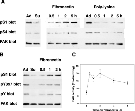
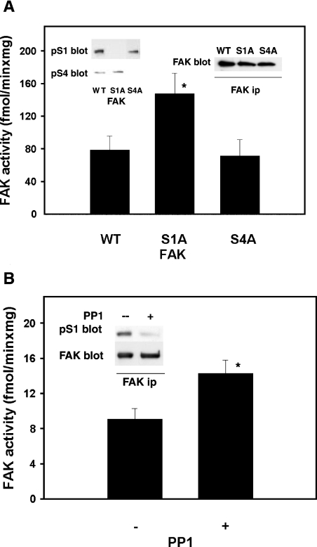
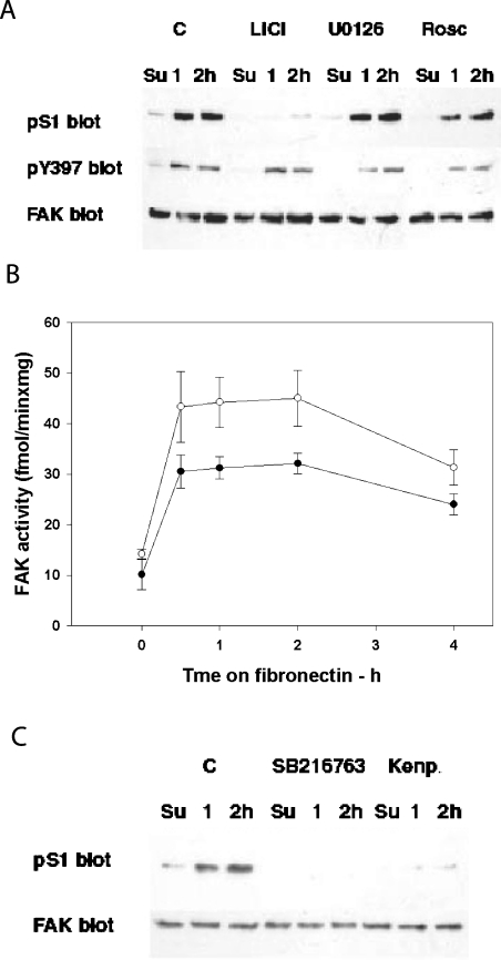
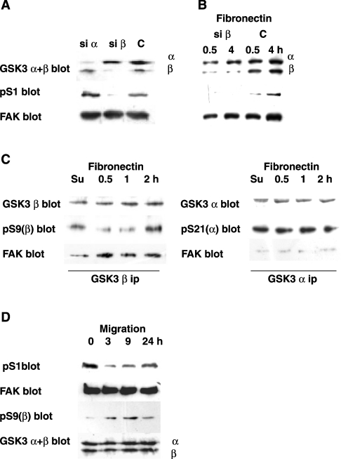
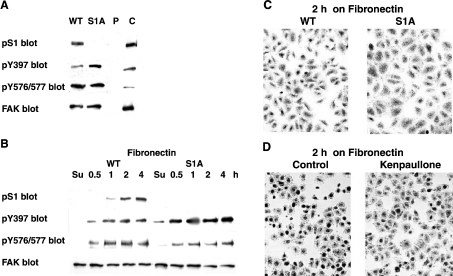
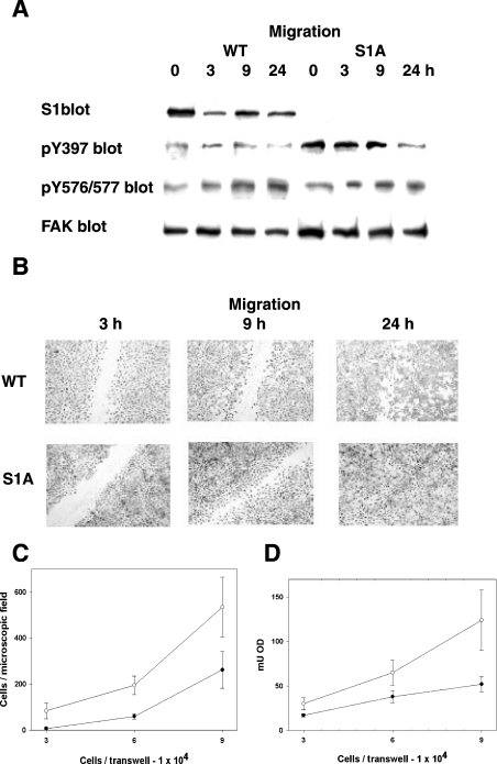
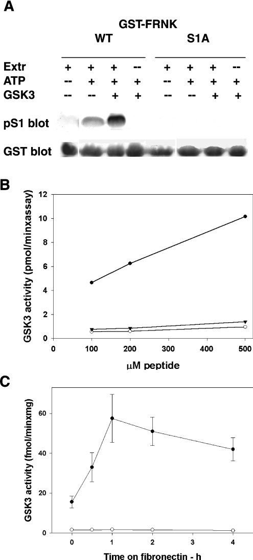
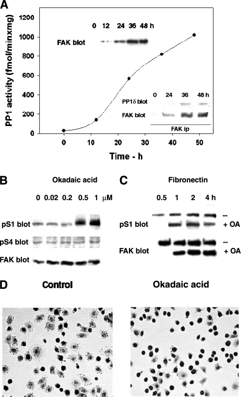
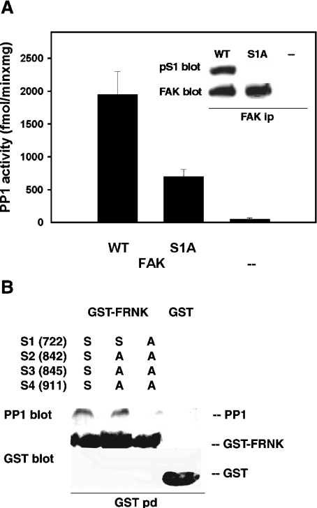
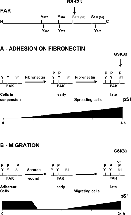
In addition to tyrosine sites, FAK (focal adhesion kinase) is phosphorylated on multiple serine residues. In the present study, the regulation of two of these sites, Ser-722 (S1) and Ser-911 (S4), was investigated. Phosphorylation of S1 (but not S4) decreased in resuspended cells, and recovered during spreading on fibronectin, indicating adhesion-dependent regulation. GSK3 (glycogen synthase kinase 3) inhibitors decreased S1 phosphorylation, and siRNA (short interfering RNA) silencing indicated further the involvement of GSK3beta. Furthermore, GSK3beta was found to become activated during cell spreading on fibronectin, and to physically associate with FAK. S1 phosphorylation was observed to decrease in wounded cell monolayers, while GSK3beta underwent inactivation and later was observed to increase to the original level within 24 h. Direct phosphorylation of S1, requiring pre-phosphorylation of Ser-726 in the +4 position, was demonstrated using purified GSK3 and a synthetic peptide containing FAK residues 714-730. An inhibitory role for S1 phosphorylation in FAK signalling was indicated by findings that both alanine substitution for S1 and dephosphorylation of S1 by PP1 (serine/threonine protein phosphatase type-1) resulted in an increase in FAK kinase activity; likewise, this role was also shown by cell treatment with the GSK3 inhibitor LiCl. The inhibitory role was confirmed by the finding that cells expressing FAK with alanine substitution for S1 displayed improved cell spreading and faster migration in wound-healing and trans-well assays. Finally, the finding that S1 phosphorylation increased in cells treated with the PP1 inhibitor okadaic acid indicated targeting of this site by PP1. These results indicate an additional mechanism for regulation of FAK activity during cell spreading and migration, involving Ser-722 phosphorylation modulated through the competing actions of GSK3beta and PP1.


Figures

Figure 1
Figure 1. FAK phosphorylation and activation during spreading on fibronectin
(A) Phosphorylation of FAK at sites S1 (Ser-722) and S4 (Ser-911 in chicken, Ser-910 in human and mouse and Ser-913 in rat FAK) during adhesion/spreading on fibronectin or polylysine. Rat fibroblasts were collected by trypsin/EDTA treatment, re-plated on dishes coated with 10 μg/ml fibronectin or 20 μg/ml polylysine and grown for up to 5 h. Lysates obtained from adherent (Ad) or resuspended (Su) fibroblasts, or from fibroblasts collected at the indicated time points were subjected to electrophoresis and Western blotting and probed separately to detect phospho-S1 (pS1), phospho-S4 (pS4) or FAK. (B) Activation of FAK during spreading on fibronectin. Cell lysates obtained as described in (A) were probed for pS1, pTyr-397 of FAK (pY397), phosphotyrosine of FAK (pY) and FAK. (C) FAK activity during spreading on fibronectin. HEK-293 cells were resuspended (at zero time), re-plated on fibronectin and collected at the indicated time points. FAK was immunoprecipitated and assayed for kinase activity using the substrate poly(Glu-Tyr). Results are shown as the means±S.E.M. for 6 separate experiments.
Figure 2
Figure 2. Activation state of wild-type and FAK mutants, and FAK activity following dephosphorylation by PP1
(A) Activation state of wild-type FAK and the S1A and S4A mutants. FAK−/− cells were transfected with a vector encoding either wild-type chicken FAK (WT) or the FAK point mutants S1A or S4A. After 24 h, cell lysates were prepared and subjected to electrophoresis, Western blotting and probing for pS4 and pS1 in sequence (left inset). FAK was immunoprecipitated (ip) from cell lysates and probed for FAK (right inset) or assayed for the protein kinase activity, as described in Figure 1(C) (bar graph). Results shown are the means±S.E.M. for 5 determinations (6 for S1A); *P≤0.05 (Student's t test) for S1A compared with WT. (B) Kinase activity of FAK treated in vitro with PP1. FAK was immunoprecipitated from rat fibroblasts in duplicate, and the immunocomplexes were incubated further with either 1 unit of PP1 catalytic subunit (purified from muscle; +) or PP1 inhibited by 0.6 μM okadaic acid (−) at 30° for 20 min and assayed for kinase activity (bar graph). Results are shown as means±S.E.M. for 5 determinations; *P≤0.05 (Student's t test). The inset shows dephosphorylation of S1 by PP1. FAK was immunoprecipitated and treated with PP1 as above. This was followed by electrophoresis, Western blotting, probing for pS1, stripping and re-probing for FAK.
Figure 3
Figure 3. FAK phosphorylation in cells treated with the inhibitors of Pro-directed kinases, and effect of LiCl on FAK activity during spreading on fibronectin
(A) Phosphorylation of S1 and Tyr-397 of FAK in cells treated with the inhibitors of Pro-directed kinases. Rat fibroblasts were plated on fibronectin in the presence of 40 mM LiCl (to inhibit GSK3), 10 μM UO126 [to inhibit MAPK (Erk-1/2)], 30 μM roscovitine (to inhibit CDKs) or DMSO as a control (C). Lysates from cells collected at the indicated time points were used to prepare three sets of immunoblots, which were then probed for pS1, pTyr-397 (pY397) and FAK. (B) Effect of LiCl on FAK activity during spreading on fibronectin. Lysates obtained from cells treated with LiCl as in (A) (○) or from untreated controls (●) were used to immunoprecipitate FAK and assay FAK kinase activity (as described in legend for Figure 1C). Results are shown as means±S.E.M. for 4 determinations. (C) Phosphorylation of S1 in cells treated with the GSK3 inhibitors SB216763 or Kenpaullone (Kenp.). Cells were treated with either 5 μM SB216763 or 10 μM Kenpaullone; other treatments were as for (A). Su, resuspended fibroblasts.
Figure 4
Figure 4. Involvement of GSK3b in FAK phosphorylation during spreading on fibronectin and migration
(A) Involvement of GSK3β in S1 phosphorylation. Rat fibroblasts were co-transfected with either the GSK3α (si α) or the GSK3β (si β) hairpin siRNA vector, together with a vector that expresses the puromycin-resistance gene. Control cells (C) were transfected with the puromycin-resistance vector only. At 24 h after transfection, the cells were exposed to 10 μg/ml puromycin to kill non-transfected cells, and collected at 72 h. Cell lysates were used to detect the GSK3 isoforms (using a mix of anti-GSK3α and β antibodies; GSK3 α+β blot), pS1 and FAK. (B) Effect of GSK3β silencing on S1 phosphorylation during spreading on fibronectin. Transfected rat fibroblasts (si β and C, as in A) were collected at 72 h and re-plated on fibronectin. Lysates obtained at 0.5 or 4 h were used to detect GSK3 α and β, pS1 and FAK. (C) GSK3β activation and association with FAK during spreading on fibronectin. Lysates from rat fibroblasts collected during spreading on fibronectin were used to immunoprecipitate GSK3β (GSK3β ip) or GSK3α (GSK3α ip). Following electrophoresis and transblotting, the blots were probed to detect GSK3β, pS9 of GSK3β [pS9(β) blot] and FAK, in sequence or GSK3α, pS21 of GSK3α [pS21(α) blot] and FAK, in sequence. (D) S1 phosphorylation in scratch-wound healing. Confluent rat fibroblasts were scratched and induced to migrate, as described in the Materials and methods section. Cells were collected at 3, 9 and 24 h from scratching, as well as from an untreated control (zero time). Lysates were subjected to electrophoresis (loading the same amounts of protein in each lane) and Western blotting. Separate blots were probed to detect pS1, FAK, pS9 of GSK3β and GSK3 α+β.
Figure 5
Figure 5. Effect of stable expression of the S1A FAK mutant or of GSK3 inhibition on cell spreading on fibronectin
(A) Stable expression of wild-type and S1A FAK in FAK−/− cells. Stable transfectants of chicken FAK were prepared and subcloned (as described further in the Materials and methods section). Cell lysates prepared from these cells, as well as from cells transfected only with the puromycin-resistance vector (P) or from rat fibroblasts (C), were used to immunodetect pS1, pTyr-397 (Y397), phospho-Y576/Y577 of FAK (pY576/577) and FAK. (B) Phosphorylation of S1, Y397 and Y576/577 during spreading on fibronectin of FAK−/− cells expressing WT or S1A FAK. Cells, as in (A), were re-plated on fibronectin and collected at the indicated time-points (as in Figure 1A). Immunodetection was as in (A). (C) FAK−/− cells expressing WT or S1A FAK at 2 h from plating on fibronectin. Cells, as in (A), were plated on fibronectin, fixed at 2 h and stained with Giemsa stain. (D) Rat fibroblasts at 2 h from plating on fibronectin in the presence of the GSK3 inhibitor Kenpaullone. Fibroblasts were plated on fibronectin in the absence (control) or in the presence of 10 μM Kenpaullone (see Figure 3), fixed and stained as in (C).
Figure 6
Figure 6. Effect of stable expression of the S1A FAK mutant on cell migration
(A) Phosphorylation of S1, Tyr-397 and Y576/577 in scratch-wound healing of FAK−/− cells expressing WT or S1A FAK. Confluent cells were induced to migrate by scratching (as in Figure 4D). The lysates of cells collected at the indicated time points were used to detect pS1, pTyr-397 (pTyr397), pY576/577 and FAK. (B) Scratch-wound healing of FAK−/− cells expressing WT or S1A FAK. Cells as in (A) were fixed at the indicated time-points and stained with Giemsa stain. (C and D) Trans-well migration assay of FAK−/− cells expressing WT or S1A FAK. The indicated amounts of cells were applied to the upper chamber of trans-well membranes, grown overnight and fixed (as detailed further in the Materials and methods section). The migrated cells were stained with Crystal Violet, counted (C) and lysed for subsequent O.D. (attenuance) determination (D).
Figure 7
Figure 7. Phosphorylation of FRNK or of a synthetic peptide by GSK3, and association of FAK and GSK3 during spreading on fibronectin
(A) Phosphorylation of S1 of FRNK by GSK3. GST–FRNK (C-terminus of FAK), either the wild-type or the S1A mutant protein, was bound to glutathione–Sepharose beads, divided into aliquots and incubated as follows: one aliquot with 0.1 mg of HeLa cell extract (Extr) and two aliquots in the presence of extract and ATP/MgCl2. One tube was incubated further with 62 m-units of purified GSK3. GSK3 and ATP/MgCl2 were also added to a remaining fourth aliquot. This was followed by electrophoresis, Western blotting, probing for pS1, stripping and re-probing for GST. (B) Phosphorylation of a peptide mimicking the FAK-S1 site by GSK3. The phosphopeptide KPSRPGYPSPRSpSEGFY (FAK residues 714–730) (●) or its mutated S1A derivative (○) were incubated with 10.3 m-units of purified GSK3, under conditions described further in the Materials and methods section. The wildtype peptide was also incubated in the presence of 50 mM LiCl (▼). Results shown are the mean values for 6 determinations. (C) FAK-associated GSK3 kinase activity during cell spreading on fibronectin, assayed with the peptide mimicking the S1 site. FAK was immunoprecipitated from rat fibroblasts at the indicated time points and assayed for the associated kinase activity with the phosphopeptide KPSRPGYPSPRSpSEGFY (FAK residues 714–730) in the absence (●) or the presence (○) of 50 mM LiCl, under conditions described further in the Materials and methods section. Results are shown as the means±S.E.M. for 4 determinations.
Figure 8
Figure 8. Association of PP1 and FAK, and FAK dephosphorylation by PP1
(A) PP1 binding to FAK during FAK induction. FAK−/− fibroblasts carrying a tetracycline-repressed FAK construct were grown in the absence of tetracycline for up to 48 h, and collected at the indicated time points. FAK was immunoprecipitated from cell lysates and assayed for the associated PP1 activity (see the Materials and methods section). Top inset: FAK expression. FAK was immunodetected in lysates from cell extracts obtained as above. Lower inset: association of PP1 and FAK. FAK was immunoprecipitated, subjected to electrophoresis and Western blotting and probed for the presence of PP1δ and FAK, after membrane stripping. (B) S1 and S4 phosphorylation in adherent cells treated with okadaic acid. Rat fibroblasts were treated with the indicated amounts of the PP1 inhibitor okadaic acid for 1 h, and the lysates were subjected to electrophoresis and immunoblotting. Two sets of samples were prepared, one to detect pS4 and FAK following membrane stripping, the second to detect pS1. (C) S1 phosphorylation in cells treated with okadaic acid during spreading on fibronectin. Rat fibroblasts were plated on fibronectin and exposed to 0.5 μM okadaic acid (+) or DMSO (−) 30 min after plating. Cells were collected as indicated and the lysates were subjected to electrophoresis and Western blotting to detect pS1 and FAK. (D) Cells at 2 h from plating on fibronectin in the presence of okadaic acid. Rat fibroblasts were plated on fibronectin in the absence (control) or in the presence of okadaic acid (as in C), fixed at 2 h from plating and stained with Giemsa stain.
Figure 9
Figure 9. PP1 binding to FAK and FRNK mutants
(A) PP1 binding to S1A FAK. FAK−/− cells were transfected with the vector encoding either wild-type (WT) or S1A FAK, as described in the legend to Figure 2. After 24 h the cells were lysed, and FAK was immunoprecipitated and assayed for the associated PP1 activity (as in Figure 8A). Results are shown as the means±S.E.M. for 3 determinations. Inset: FAK was immunoprecipitated from cells expressing the indicated FAK type or the empty vector (−), and then subjected to electrophoresis, Western blotting and probing for pS1 and FAK. (B) PP1 binding to FRNK point mutants in vitro. The indicated serine-to-alanine mutants of S1 (Ser-722), S2 (Ser-842), S3 (Ser-845) and S4 (Ser-911) of FRNK, or GST alone, were bound to glutathione–Sepharose beads and used to pull-down purified PP1 catalytic subunit, which was then detected by immunoblotting (PP1 blot). This was followed by stripping and detection of FRNK mutants or GST (GST blot).
Figure 10
Figure 10. FAK scheme depicting the major tyrosine and serine phosphorylation sites
S1 phosphorylation by GSK3β and changes in S1 phosphorylation during cell adhesion on fibronectin and cell migration are shown.

Similar articles

Cited by

References

    1. Hanks S. K., Ryzhova L., Shin N., Brabek J. Focal adhesion kinase signalling activities and their implications in the control of cell survival and motility. Front. Biosci. 2003;8:982–986. - PubMed
    1. Parsons J. T. Focal adhesion kinase: the first ten years. J. Cell Sci. 2003;116:1409–1416. - PubMed
    1. Carragher N. O., Frame M. C. Focal adhesion and actin dynamics: a place where kinases and proteases meet to promote invasion. Trends Cell Biol. 2004;14:241–249. - PubMed
    1. Owen J. D., Ruest P. J., Fry D. W., Hanks S. K. Induced focal adhesion kinase (FAK) expression in FAK-null cells enhances cell spreading and migration requiring both auto- and activation loop phosphorylation sites and inhibits adhesion-dependent tyrosine phosphorylation of Pyk2. Mol. Cell. Biol. 1999;19:4806–4818. - PMC - PubMed
    1. Malik R. K., Parsons J. T. Integrin-mediated signaling in normal and malignant cells: a role of protein tyrosine kinases. Biochim. Biophys. Acta. 1996;1287:73–76. - PubMed

Publication types

MeSH terms

Substances