. 2005 Sep;16(9):3999-4012.
doi: 10.1091/mbc.e05-02-0093. Epub 2005 Jun 22.

Alanine scanning of Arp1 delineates a putative binding site for Jnm1/dynamitin and Nip100/p150Glued

Affiliations

Alanine scanning of Arp1 delineates a putative binding site for Jnm1/dynamitin and Nip100/p150Glued

Sean W Clark et al. Mol Biol Cell. 2005 Sep.

Abstract

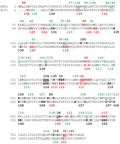
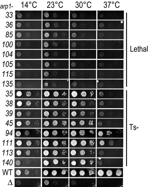
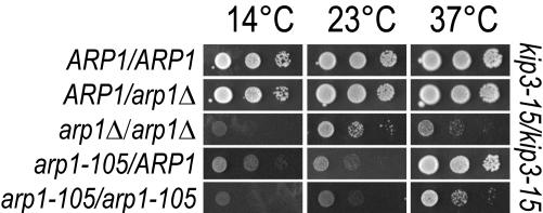
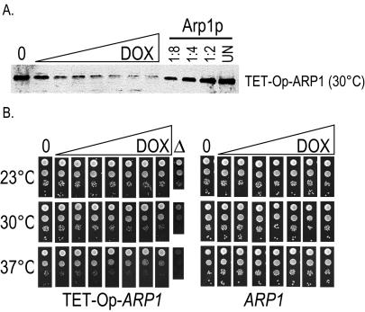
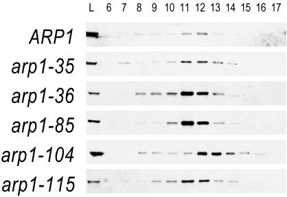
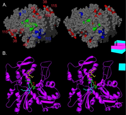
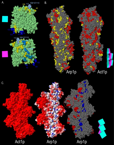
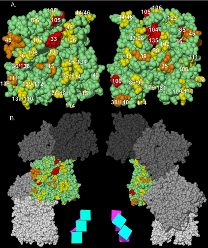
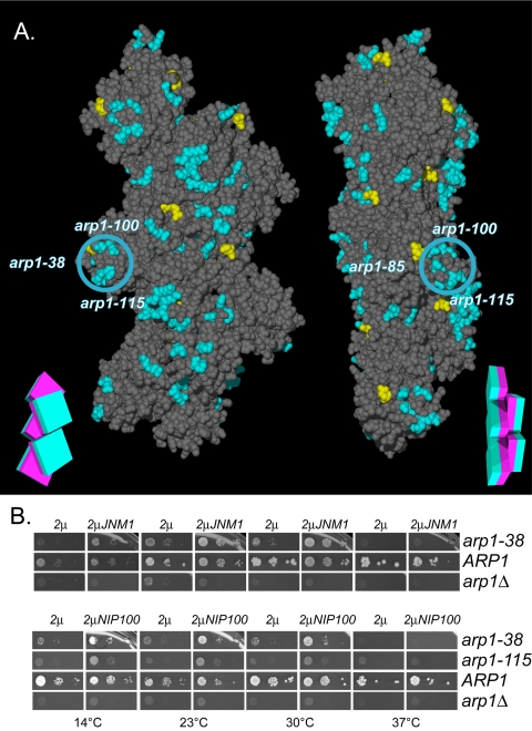
Arp1p is the only actin-related protein (ARP) known to form actin-like filaments. Unlike actin, Arp1p functions with microtubules, as part of the dynein regulator, dynactin. Arp1p's dissimilar functions imply interactions with a distinct set of proteins. To distinguish surface features relating to Arp1p's core functions and to identify the footprint of protein interactions essential for dynactin function, we performed the first complete charge-cluster-to-alanine scanning mutagenesis of an ARP and compared the results with a similar study of actin. The Arp1p mutations revealed three nonoverlapping surfaces with distinct genetic properties. One of these surfaces encompassed a region unique to Arp1p that is crucial for Jnm1p (dynamitin/p50) and Nip100p (p150(Glued)) association as well as pointed-end associations. Unlike the actin mutations, none of the ARP1 alleles disrupt filament formation; however, one pointed-end allele delayed the elution of Arp1p on gel filtration, consistent with loss of additional subunits.


Figures

Figure 1.
Figure 1.
Comparison of ARP1 and ACT1 charge-cluster to alanine mutations. ARP1 allele numbers are indicated. Green indicates pseudo-wild-type, red Ts, and black lethal. Alleles separated by a slash are overlapping charge clusters where the double underline designates the shared residues. Alanine scanning alleles for actin and their phenotypes are indicated on the lower strand. Actin data are from Wertman et al. (1992).
Figure 2.
Figure 2.
Phenotypic profiles of lethal and Ts ARP1 alleles. All 45 ARP1 alleles were examined for growth phenotypes in the kip3-15 background. Ts and lethal are shown here. Each allele was covered by a plasmid harboring wild-type ARP1 that was counterselected with 5-FOA at four temperatures. The slight growth at 23°C for arp1Δkip3Δ is typical of this strain background. Each spot shows the growth of successive, serial 1:10 dilutions from left to right. WT, ARP1+; Δ, arp1Δ.
Figure 3.
Figure 3.
Hemizygous ARP1 and the partial dominant cold sensitivity of arp1-105. Diploid strains harbored the indicated ARP1 alleles in a homozygous kip3-15 background. Hemizygous ARP1 diploids were compared with the homozygous ARP1 wild-type and homozygous arp1Δ diploids. The hemizygous configuration of ARP1 does not affect growth. These were further compared with arp1-105 in homozygous and heterozygous configurations. The decreased growth of heterozygous arp1-105 at 14 and 23°C relative to heterozygous arp1Δ indicates a dominant cold sensitivity for arp1-105. Growth was examined at three temperatures by 5-FOA counterselection of a plasmid harboring ARP1. Tenfold serial dilutions are shown.
Figure 4.
Figure 4.
(A) ARP1 under control of the tetracycline operator allows reduction of the steady-state level of wild-type Arp1p. Immunoblot of ARP1 kip3Δ strains wherein the endogenous ARP1 promoter had been replaced with a tetracycline operator and minimal promoter (TET-Op-ARP1). Cultures were grown with increasing concentrations of the tetracycline analog doxycycline (DOX) (0, no doxycycline). For comparison, serial dilutions of a wild-type ARP1 strain are shown. UN, undiluted. Arp1p was detected with anti-Arp1p antibody. (B) ARP1 under the tetracycline operator defines a growth threshold that can be equated with a steady-state level of wild-type Arp1p. Tenfold serial dilutions of a TET-Op-ARP1 kip3Δ strain and an ARP1 kip3Δ strain were examined for growth at three temperatures on solid media containing increasing concentrations of doxycycline, matching those shown in A. A growth defect is first visible between the third and fourth dilutions of doxycycline. An arp1Δ strain grown in the absence of doxycycline is shown for comparison.
Figure 5.
Figure 5.
Elution profiles of Arp1p mutants when overexpressed. Strains harboring mutant Arp1p were examined by gel filtration and Arp1p was detected by immunoblot with anti-Arp1p antibody. Fraction size is 1 ml. Under these conditions, we are likely examining only the Arp1p filament and capping subunits (see main text). The panels compare elution of wild-type Arp1p with that of Ts and lethal alleles, overexpressed from a 2μ plasmid. For wild-type, 10-fold less protein was examined. The Ts of arp1-35 and the lethal allele arp1-115 are fully rescued under these conditions, whereas the lethal allele arp1-36 is not rescued by expression to wild-type steady-state levels. Despite these phenotypic differences, all show the same peak elution. Lethal allele arp1-104, which is partially rescued by overexpression, has a notably delayed elution compared with the other mutants. Cytosols were prepared from strains transformed with a 2μ plasmid carrying the same allele as integrated at ARP1. Calibration standards were Void, 6.8 ml, thyroglobulin (669 kDa), 11.0 ml; ferritin (440 kDa), 13.0 ml; aldolase (158 kDa), 14.3 ml; ovalbumin (43 kDa), 15.6 ml; and acetone, 19.7 ml.
Figure 6.
Figure 6.
The pointed-end interface and the ATP binding pocket of Arp1p. (A) A stereoview of the pointed-end of an Arp1p pentamer. Ts and lethal residues are colored by mass-action rescue (red, rescued; blue, not rescued), and numbered by allele. arp1-104 (a rescued lethal allele) is highlighted in green. (B) Three of the ARP1 mutations line the ATP binding pocket of Arp1p. A stereoview is shown of the Arp1p ribbon structure. The ATP is represented in green. The backbone and side chains of arp1-33 are shown in yellow, arp1-101 in red, and arp1-113 in blue. A schematic representation of the filament consisting of boxes representing each monomer, each with a magenta inner face and teal outer face, serves as an orienting guide to the relative rotation of each filament.
Figure 7.
Figure 7.
The functional surfaces of the Arp1p filament. (A) Homotypic contact regions in the Arp1p filament. A single Arp1p monomer (green) makes contacts with four surrounding monomers. The charge clusters belonging to the polypeptide chain of the central monomer that make contact with surrounding monomers are shown in yellow. The side chains of charge clusters from the four adjacent monomers that contact the central monomer are shown in four shades of blue (only the relevant side chains are shown, the rest of the polypeptide is not shown for clarity). Opposite faces of the central monomer are shown. The schematic magenta box represents the monomer's inner facing surface; the teal box represents the outer face. (B) The Arp1p filament pseudo-wild-type surface. ARP1 and ACT1 alleles are mapped onto a section of their respective filaments. The “pointed-end” is up and the “barbed-end” is down in all images. Although likely shorter than the actual Arp1p filament, a pentamer is shown to allow visualization of trends, while still providing sufficient detail. Red indicates Ts and lethal alleles; yellow indicates pseudo-wild type. (C) The positive-charge and mass-action rescue surfaces of Arp1p. We compared the electrostatic potential of Act1p and Arp1p pentamers. Arp1 has a net positive charge surface that encompasses the bulk of Ts and lethal alleles. Red, –3.5; blue, 3.5 (bottom, left and middle filaments). For mass-action rescue, red, rescued, and blue, nonrescued (bottom, right filament). Note the coincident position of the positive-charge surface and the nonrescued alleles. For clarity, pseudo-wild-type alleles are not shown; they would be found along the three projections to the right. The red circle indicates the Arp1-specific projection encompassing arp-38, 100, and 115. A schematic representation of the Arp1p filament consisting of five boxes, each with a magenta inner face and teal outer face, serves as an orienting guide to the relative position of monomers in each filament.
Figure 8.
Figure 8.
ARP1 alleles mapped on a predicted structure of the Arp1p filament. (a) Alleles are represented by number. arp1-94 is not indicated because it resides in the extreme N terminus, not represented in the structure. The “pointed-end” is up and the “barbed-end” is down in all images. Yellow indicates pseudo-wild type, orange indicates Ts, and red indicates Lethal. (b) The monomers in (a) are shown relative to the Arp1p filament. A schematic representation of the filament consisting of five boxes, each with a magenta inner face and teal outer face, serves as an orienting guide to the relative rotation of each filament.
Figure 9.
Figure 9.
Jnm1–Arp1p 2-hybrid interaction and overexpression-rescue of the Arp1p-specific arp1-100/115 projection by Jnm1p and Nip100p. (A) The results of two-hybrid interaction between Jnm1p and mutant Arp1p. Mutation of the Arp1p residues shown in blue decreased the interaction with Jnm1p, those in yellow had no effect. The blue circle indicates the position of the Arp1p-specific projection, encompassing arp1-38, 100, and 115. A schematic representation of the filament consisting of five boxes, each with a magenta inner face and teal outer face, serves as an orienting guide to the relative rotation of each filament. (B) Plasmids overexpressing (2μ His+) either JNM1 or NIP100 were introduced into all Ts and lethal ARP1 mutant strains, ARP1 wild-type, or arp1Δ. Their ability to rescue the ARP1 mutation was accessed by growth under conditions (5-FOA-HIS) that counterselect the wild-type ARP1 covering plasmid. Though all Ts and lethal alleles were examined, only the ARP1 alleles demonstrating rescue are shown.

Similar articles

Cited by

References

    1. Amberg, D. C., Basart, E., and Botstein, D. (1995). Defining protein interactions with yeast actin in vivo. Nat. Struct. Biol. 2, 28–35. - PubMed
    1. Belli, G., Gari, E., Aldea, M., and Herrero, E. (1998a). Functional analysis of yeast essential genes using a promoter-substitution cassette and the tetracycline-regulatable dual expression system. Yeast 14, 1127–1138. - PubMed
    1. Belli, G., Gari, E., Piedrafita, L., Aldea, M., and Herrero, E. (1998b). An activator/repressor dual system allows tight tetracycline-regulated gene expression in budding yeast. Nucleic Acids Res. 26, 942–947. - PMC - PubMed
    1. Belmont, L. D., and Drubin, D. G. (1998). The yeast V159N actin mutant reveals roles for actin dynamics in vivo. J. Cell Biol. 142, 1289–1299. - PMC - PubMed
    1. Belmont, L. D., Orlova, A., Drubin, D. G., and Egelman, E. H. (1999a). A change in actin conformation associated with filament instability after Pi release. Proc. Natl. Acad. Sci. USA 96, 29–34. - PMC - PubMed

Publication types

MeSH terms