. 2005 Sep;16(9):4124-38.
doi: 10.1091/mbc.e04-11-0976. Epub 2005 Jun 22.

The novel fission yeast protein Pal1p interacts with Hip1-related Sla2p/End4p and is involved in cellular morphogenesis

Affiliations

The novel fission yeast protein Pal1p interacts with Hip1-related Sla2p/End4p and is involved in cellular morphogenesis

Wanzhong Ge et al. Mol Biol Cell. 2005 Sep.

Abstract

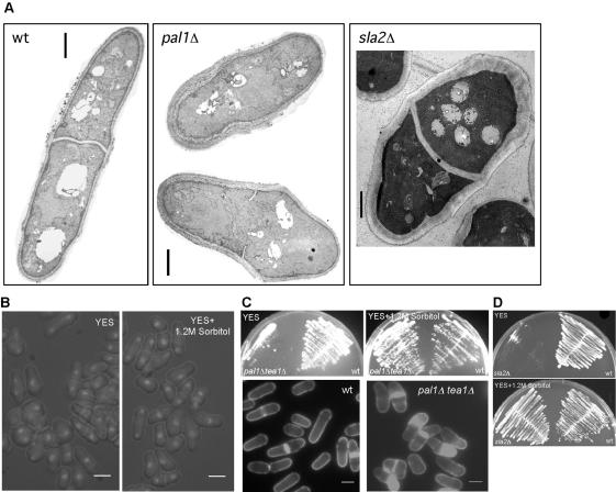
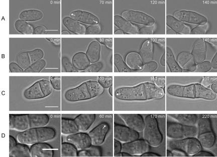
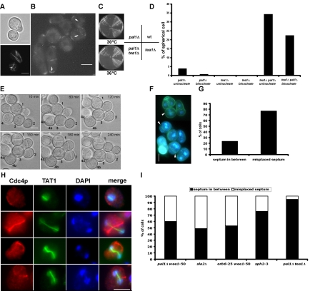
The establishment and maintenance of characteristic cellular morphologies is a fundamental property of all cells. Here we describe Schizosaccharomyces pombe Pal1p, a protein important for maintenance of cylindrical cellular morphology. Pal1p is a novel membrane-associated protein that localizes to the growing tips of interphase cells and to the division site in cells undergoing cytokinesis in an F-actin- and microtubule-independent manner. Cells deleted for pal1 display morphological defects, characterized by the occurrence of spherical and pear-shaped cells with an abnormal cell wall. Pal1p physically interacts and displays overlapping localization with the Huntingtin-interacting-protein (Hip1)-related protein Sla2p/End4p, which is also required for establishment of cylindrical cellular morphology. Sla2p is important for efficient localization of Pal1p to the sites of polarized growth and appears to function upstream of Pal1p. Interestingly, spherical pal1Delta mutants polarize to establish a pearlike morphology before mitosis in a manner dependent on the kelch-repeat protein Tea1p and the cell cycle inhibitory kinase Wee1p. Thus, overlapping mechanisms involving Pal1p, Tea1p, and Sla2p contribute to the establishment of cylindrical cellular morphology, which is important for proper spatial regulation of cytokinesis.


Figures

Figure 1.
Figure 1.
Identification of Pal1p in fission yeast. (A) Schizosaccharomyces pombe Pal1p is aligned with S. cerevisiae YDR348cp, Magnaporthe grisea MG02779.4, Neurospora crassa NCU00687.1. Identical amino acids are shaded in black and similar amino acids are shaded in gray. (B) Schematic representation of the homology between Sp.Pal1p and other related proteins. Identities (%) and the number of amino acids in parentheses are indicated in the conserved region. (C) Localization of Pal1p in wild-type cells. Cells expressing functional GFP-tagged Pal1p grown on YES plates were visualized by fluorescence microscopy. Pal1p localizes to the growing end(s) during monopolar and bipolar growth (cells 1 and 2) and to the medial cell division site during cytokinesis (cells 3–6). Scale bar, 5 μm. (D) Detection of Pal1p. Total cell extracts were prepared from the pal1-GFP (lane 1) and wild-type (lane 2) strains and were subjected to SDS-PAGE and immunoblotted using α-GFP antibody. (E) Pal1p is a membrane associated protein. Total cell extracts prepared from the pal1-GFP strain were fractionated by density gradient centrifugation. The fractions obtained were subjected to SDS-PAGE and immunoblotted using α-GFP, α-PMA1, and α-Cdc8p antibodies. Lanes 1–6 show the different fractions from top to bottom of the density gradient.
Figure 2.
Figure 2.
Pal1p localizes to the sites of cell growth and division in an F-actin- and microtubule-independent manner. Different strains with functional GFP-tagged Pal1p were grown on YES agar plate at 24°C and then shifted to 36°C for 6 h. (A) orb2-34 at 24°C; a shows Pal1p-GFP; b shows aniline blue staining; (B) cdc25-22 at 36°C; (C) orb3-167 at 36°C; (D) tea1Δ; (E) mid1Δ; (F) Pal1p localizes to the zone of cell fusion in the h90 strain. (G) Role of F-actin in Pal1p localization. cdc25-22 pal1-GFP cells were blocked for 4 h at 36°C in YES medium and then released to 24°C into medium containing 15 μM Lat A or DMSO. Cell samples were taken at different time points (a, 60 min; b, 60 min; c, 120 min; d, 180 min) after release and visualized by fluorescence microscopy. (H) Role of microtubules in Pal1p localization. nda3-KM311 pal1-GFP cells were grown on a YES agar plate at 32°C for 1 d and shifted to 19°C for 6 h. Scale bar, 5 μm.
Figure 3.
Figure 3.
Pal1p physically interacts with Sla2p, a Hip1-related protein in S. pombe. (A) Schematic representation of the homology between fission yeast Sla2p and other Sla2p/Hip1p family proteins. S. pombe Sla2p (Sp_Sla2p) is aligned with S. cerevisiae Sla2p (Sc_Sla2p), Homo sapiens Hip1p (Hs_Hip1p), Homo sapiens Tln1p (Hs_Tln1p). Identities (%) and the number of amino acids are indicated in two conserved domains, ANTH and I/LWEQ. (B) Coimmunoprecipitation of Pal1p with Sla2p in α-GFP immunoprecipitation from native cell extracts. Total cell extracts were prepared from pal1-Myc, sla2-GFP, pal1-Myc sla2-GFP, pal1-GFP, sla2-Myc, and pal1-GFP sla2-Myc strains. The entire immune complex prepared from 500 μl of lysate and 30 μl of straight-lysate were subjected to SDS-PAGE and immunoblotted with α-GFP or α-Myc antibodies. (C) Localization of Sla2p-GFP in wild-type cells. Cells expressing functional Sla2p-GFP under its native promoter were grown at 30°C and visualized by fluorescence microscopy. (D) Colocalization of Sla2p-GFP and F-actin patches. Cells expressing Sla2p-GFP were grown at 30°C and immunostained with α-GFP antibody and Alexa Fluor-488-conjugated phalloidin. (E) Role of F-actin in Sla2p localization. cdc25-22 sla2-GFP cells were blocked at 36°C for 4 h in YES medium and then released to 24°C into YES medium containing 15 μM Lat A or DMSO. Cell samples were taken at different time points (a, 60 min; b, 60 min; c, 120 min; d, 180 min) after release. (F) Role of microtubules in Sla2p localization. nda3-KM311 sla2-GFP cells were grown in YES liquid medium at 32°C and then shifted to 19°C for 6 h. Scale bar, 5 μm.
Figure 4.
Figure 4.
pal1Δ and sla2Δ cells have defects in cell morphology and cell wall. (A) Microscopic analysis of pal1Δ cells. pal1Δ cells were grown in minimal medium at 30°C and stained with DAPI to visualize DNA. Arrows, spherical cell; arrowheads, abnormal shaped cell. (B) pal1Δ cells were grown in minimal medium, fixed, and stained with Alexa Fluor-488-conjugated phalloidin to show the F-actin localization. (C) pal1Δ cells expressing GFP-α-tubulin were used to visualize interphase microtubules. (D) Wild-type and pal1Δ cells were grown in YES liquid medium and stained with Calcofluor. (E) Morphology of sla2Δ cells. sla2Δ cells were grown in YES supplemented with 1.2 M sorbitol, washed, and reinoculated into YES medium for 6 h at 30°C, fixed, and stained with aniline blue and DAPI to visualize division septa and nuclei, respectively. Scale bar, 5 μm.
Figure 5.
Figure 5.
pal1Δ and sla2Δ cells have abnormally thick cell wall and are rescued by growth on sorbitol. (A) Exponentially growing wild-type, pal1Δ, and sla2Δ cells were processed for thin section electron microscopy. Electron micrographs of wild-type, pal1Δ cells, and sla2Δ cells are shown in the left, middle, and right panels, respectively. Scale bar, 1 μm. (B) Morphology of pal1Δ mutants is suppressed by sorbitol. pal1Δ cells were grown in YES and YES plus 1.2 M sorbitol medium. Cells were stained with DAPI and visualized by fluorescence microscopy. (C) pal1Δ phenotype can be suppressed by sorbitol in pal1Δ tea1Δ mutant. pal1Δ tea1Δ and wild-type cells were grown on YES plate with or without 1.2 M sorbitol for 2 d at 36°C (top panel). Cells from the plates were stained with aniline blue to visualize the cell shape (bottom panel). (D) Ability of sla2Δ cells to form colonies on YES supplemented with sorbitol. Wild-type and sla2Δ cells were streaked on YES plates with or without sorbitol and incubated at 32°C for 3 d. Scale bar, 5 μm.
Figure 6.
Figure 6.
Dependency relationships between Sla2p and Pal1p. (A) Cells expressing functional GFP-tagged Sla2p in wild-type (a) and pal1Δ (b) backgrounds were grown in YES liquid medium and imaged by laser scanning confocal microscopy. Cells expressing functional GFP-tagged Pal1p in wild-type (c) and sla2Δ (d) backgrounds were grown in YES liquid medium supplemented with sorbitol and imaged by laser scanning confocal microscopy. Scale bar, 5 μm. (B) Viability of sla2Δ cells at high temperature is suppressed by multiple copies of pal1. sla2Δ cells carrying pal1 or sla2 plasmid were streaked on minimal medium without leucine supplement and were grown at 24°C (a) and 36°C (b) for 3 d. sla2Δ transformed with the empty vector, pTN-L1, was included as a control. (C) Microscopic analysis of sla2Δ cells transformed with vector (a), sla2 (b), and pal1 (c) plasmids. sla2Δ cells carrying vector, sla2, and pal1 plasmids were grown in minimal medium without leucine supplement at 24°C, shifted to 36°C for 8 h, and then stained with Calcofluor. Scale bar, 5 μm.
Figure 7.
Figure 7.
Growth patterns of pal1Δ cells. pal1Δ cells (>5 for each morphology) were imaged by time-lapse light microscopy on minimal medium agar pads at room temperature. (A) Bipolar growth pattern of a cylindrical cell. (B) Pattern of growth of the spherical and cylindrical cell generated by division of a pear-shaped cell. Note that the spherical cell generates a tip at or near the new end. (C) Pattern of growth of cells defective in separation. Note the old end growth and no new end (branching) growth. (D) Pattern of growth of pear-shaped cells generated by cell division. Note that both daughters carry out old end growth. Scale bar, 5 μm.
Figure 8.
Figure 8.
Establishment of cylindrical morphology in spherical cells of pal1Δ. (A) Correlation of cell shape and nuclear number in pal1Δ cells. Cells were grown in minimal medium at 30°C to log phase, fixed, and stained with DAPI to visualize DNA. At least 600 cells each were counted for uninucleate and binucleate categories. (B) Time-lapse analysis of the growth of pal1Δ mutant. Cells were imaged by time-lapse light microscopy on minimal medium agar pads at room temperature. Time in minutes is indicated on the top right of each panel at the commencement of observation (t = 0). Cells are numbered for tracking, a and b are used to indicate the products of cell division. (C) Spherical shaped pal1Δ cells repolarize while the Cdc13-YFP signal is present in the nucleus. pal1Δ cells expressing Cdc13-YFP were imaged by time-lapse laser scanning confocal microscopy on YES medium agar pads at room temperature. (D) Wee1p is required for the viability and the repolarization of pal1Δ mutant. Wild-type, pal1Δ, wee1-50, and pal1Δ wee1-50 were streaked on YES agar plate and incubated for 2 d at 36°C. (E) Microscopic analysis of wee1-50 and pal1Δ wee1-50 mutants. Cells were stained with DAPI to show DNA. (F) Quantification of spherical cells with one or two nuclei in pal1Δ, wee1-50, and pal1Δ wee1-50 mutants. Cultures were grown in YES medium at 24°C to log phase and shifted to 36°C for 4 h. Cell samples were stained with DAPI and at least 300 cells were counted for each strain. Scale bar, 5 μm.
Figure 9.
Figure 9.
Tea1p is essential for the viability and repolarization of pal1Δ cells. (A) Microtubules converge into the cylindrical projection during the repolarization of pal1Δ cells. pal1Δ cells expressing α-tubulin GFP were grown on minimal medium agar pad at room temperature and imaged by confocal microscopy. (B) Tea1p-YFP is spread over the cortex in spherical pal1Δ cells and localizes at tips generated in spherical pal1Δ cells. pal1Δ cells expressing Tea1p-YFP were used to visualize the localization of Tea1p-YFP. (C) Wild-type, pal1Δ, tea1Δ, and pal1Δ tea1Δ were streaked on YES agar plate and incubated for 2 d at 30 or 36°C. (D) Quantification of the percentages of spherical cells with one or two nuclei in pal1Δ, tea1Δ, and pal1Δ tea1Δ mutants. Cultures were grown in YES medium at 24°C to log phase and shifted to 36°C for 6 h. Cell samples were stained with DAPI and at least 300 cells were counted for each strain. (E) The growth of pal1Δ tea1Δ was imaged by time-lapse light microscopy on YES agar pads at room temperature. Time is indicated in minutes from the beginning of observation (t = 0). Cells are numbered for tracking, a and b are used to indicate the daughter cells produced after cell division. (F) Microscopic analysis of pal1Δ tea1Δ cells. Cells were stained with DAPI and aniline blue to visualize DNA and septum. Arrowheads indicate the septum. (G) Quantification of the percentages of spherical cells with properly positioned septum in between two nuclei or misplaced septum. (H) Localization of Cdc4p in pal1Δ tea1Δ double mutant. Exponentially growing cells were fixed with formaldehyde, processed for immunofluorescence, and stained with DAPI to visualize DNA, α-Cdc4p to visualize actomyosin ring, and α-TAT1 to visualize microtubules. Merge shows that the mitotic spindle (green) is not aligned properly with respect to the Cdc4p ring (red). (I) Septum is misplaced in different spherical mutants. pal1Δ wee1-50 and orb6-25 wee1-50 mutants were grown in YES medium at 24°C and shifted to 36°C for 5 h. sph2-3 cells were cultured in YES medium at 30°C. sla2Δ and pal1Δ tea1Δ mutants were grown in YES supplemented with 1.2 M sorbitol at 24°C and shifted to 36°C for 5 h. Scale bar, 5 μm. Except for sorbitol-grown pal1Δ tea1Δ, in which abnormal cells were counted, the phenotype in spherical cells is indicated.

Similar articles

Cited by

References

    1. Arellano, M., Duran, A., and Perez, P. (1996). Rho 1 GTPase activates the (1–3)beta-D-glucan synthase and is involved in Schizosaccharomyces pombe morphogenesis. EMBO J. 15, 4584-4591. - PMC - PubMed
    1. Arellano, M., Niccoli, T., and Nurse, P. (2002). Tea3p is a cell end marker activating polarized growth in Schizosaccharomyces pombe. Curr. Biol. 12, 751-756. - PubMed
    1. Bahler, J., and Pringle, J. R. (1998). Pom1p, a fission yeast protein kinase that provides positional information for both polarized growth and cytokinesis. Genes Dev. 12, 1356-1370. - PMC - PubMed
    1. Balasubramanian, M. K., Helfman, D. M., and Hemmingsen, S. M. (1992). A new tropomyosin essential for cytokinesis in the fission yeast S. pombe. Nature 360, 84-87. - PubMed
    1. Balasubramanian, M. K., McCollum, D., and Gould, K. L. (1997). Cytokinesis in fission yeast Schizosaccharomyces pombe. Methods Enzymol. 283, 494-506. - PubMed

Publication types

MeSH terms

LinkOut - more resources