Comparative Study
. 2005 Jun 22;25(25):5935-42.
doi: 10.1523/JNEUROSCI.1364-05.2005.

Sexual dimorphism of short-wavelength photoreceptors in the small white butterfly, Pieris rapae crucivora

Affiliations
Comparative Study

Sexual dimorphism of short-wavelength photoreceptors in the small white butterfly, Pieris rapae crucivora

Kentaro Arikawa et al. J Neurosci. .

Abstract

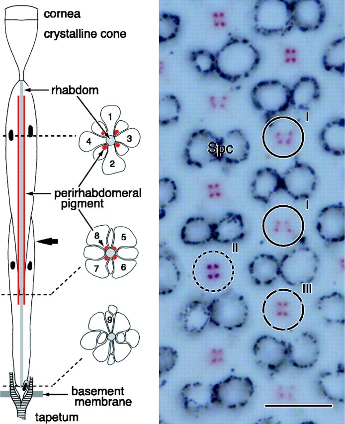
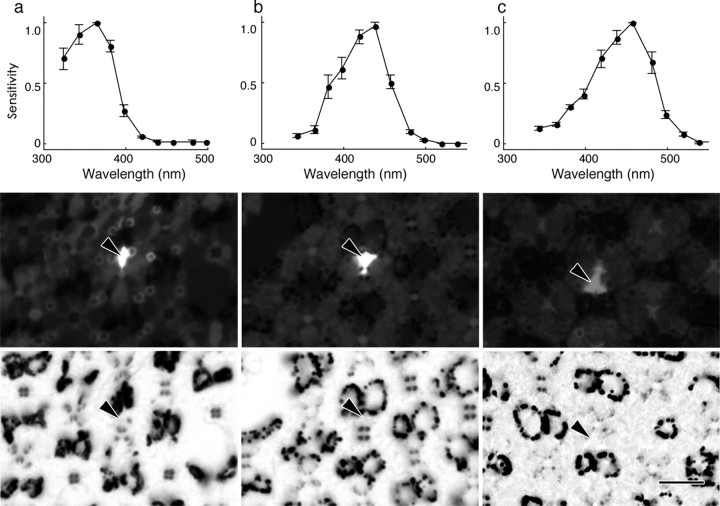
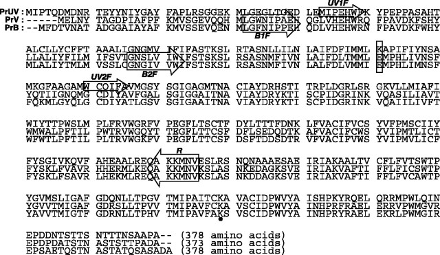
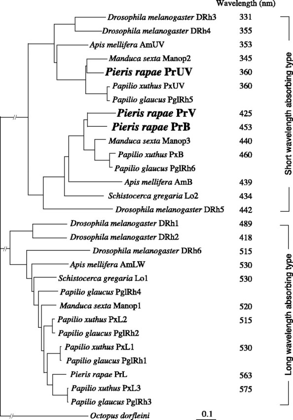
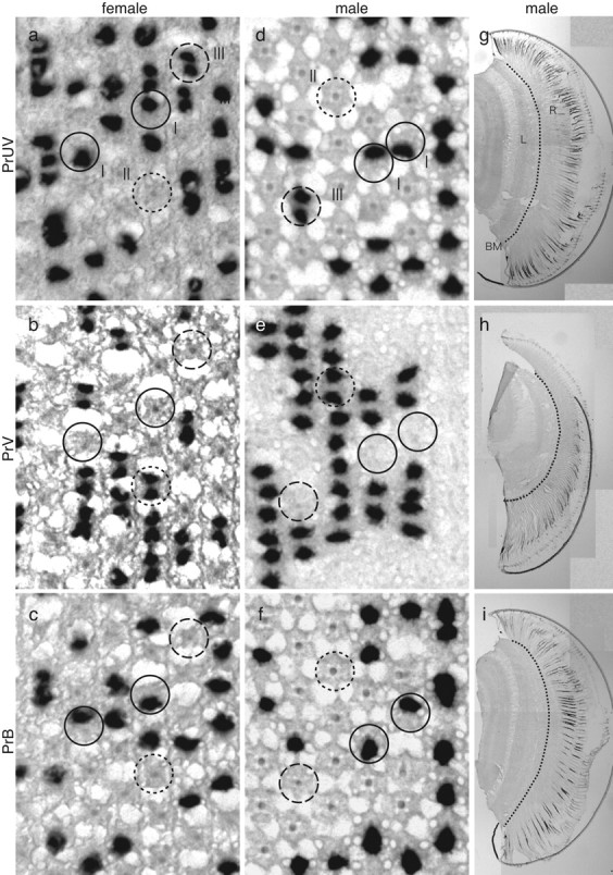
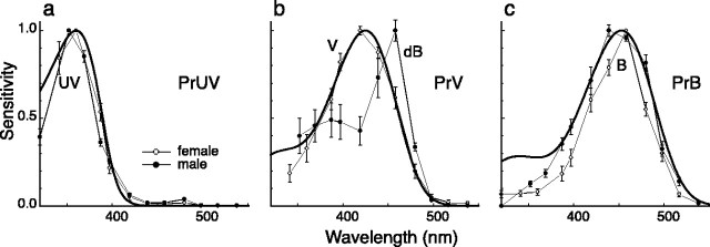
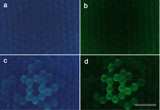
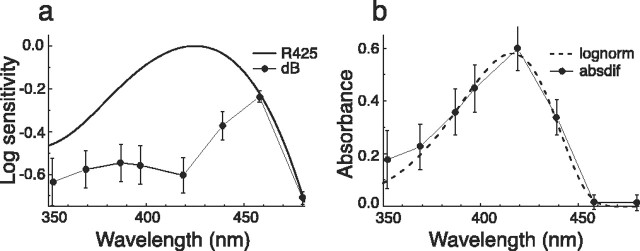
The eyes of the female small white butterfly, Pieris rapae crucivora, are furnished with three classes of short-wavelength photoreceptors, with sensitivity peaks in the ultraviolet (UV) (lambda(max) = 360 nm), violet (V) (lambda(max) = 425 nm), and blue (B) (lambda(max) = 453 nm) wavelength range. Analyzing the spectral origin of the photoreceptors, we isolated three novel mRNAs encoding opsins corresponding to short-wavelength-absorbing visual pigments. We localized the opsin mRNAs in the retinal tissue and found that each of the short-wavelength-sensitive photoreceptor classes exclusively expresses one of the opsin mRNAs. We, accordingly, termed the visual pigments PrUV, PrV, and PrB, respectively. The eyes of the male small white butterfly also use three classes of short-wavelength photoreceptors that equally uniquely express PrUV, PrV, and PrB. However, whereas the spectral sensitivities of the male photoreceptors with PrUV and PrB closely correspond to those of the female, the male photoreceptor expressing PrV has a double-peaked blue (dB) spectral sensitivity, strongly deviating from the spectral sensitivity of the female V photoreceptor. The male eyes contain a pigment that distinctly fluoresces under blue-violet as well as UV excitation light. It coexists with the dB photoreceptors and presumably acts as a spectral filter with an absorbance spectrum peaking at 416 nm. The narrow-band spectral sensitivity of the male dB photoreceptors probably evolved to improve the discrimination of the different wing colors of male and female P. rapae crucivora in the short-wavelength region of the spectrum.


Figures

Figure 1.
Figure 1.
Anatomy of an ommatidium of the female P. rapae crucivora in the main frontoventral part of the compound eye. Left, The schematic diagram shows that an ommatidium contains nine photoreceptor cells R1-R9 (1-9), with four distal photoreceptors, R1-R4, four proximal photoreceptors, R5-R8, and one basal photoreceptor, R9. The three transverse views are from the distal, proximal, and basal levels (dotted lines) of the ommatidium. The rhabdomeres, the photoreceptor organelles that contain the visual pigment, are joined into the fused rhabdom. The rhabdom is surrounded by four clusters of pigment that exist in the distal extension of the soma of the proximal photoreceptors. The bold arrow indicates the level at which the micrograph on the right was taken. Right, Depending on the ommatidium, the cluster pattern is a trapezoid (I), square (II), or rectangle (III). The pigment is pale red in ommatidial types I and III and deep red in type II. Spc, Secondary pigment cells that optically isolate the ommatidia. Scale bar, 10 μm.
Figure 2.
Figure 2.
Spectral sensitivity of short-wavelength photoreceptors in female P. rapae crucivora and their localization (arrowheads). a, A UV photoreceptor, peaking at 360 nm (top), identified as the R1 (middle) of a type I ommatidium (bottom). b, A V photoreceptor, peaking at 420 nm (top), identified as the R1 (middle) of a type II ommatidium (bottom). c, A B photoreceptor, peaking at 460 nm (top), identified as the R2 (middle) of a type I ommatidium (bottom). The spectral sensitivity curves are the means (±SE) of six (a), four (b), and four (c) consecutive measurements from single photoreceptors. Scale bar, 10 μm.
Figure 3.
Figure 3.
Deduced amino acid sequences of the three short-wavelength opsins: PrUV, PrV, and PrB. Nucleotide sequences are available in the DNA Data Bank of Japan(accession numbers: PrUV, AB208673; PrV, AB208674; PrB, AB208675). The boxed lysine and glutamate residues correspond to the sites responsible for short-wavelength-absorbing characteristics (Salcedo et al., 2003). The boxed arrows indicate regions on which forward (UV1F, UV2F, B1F, B2F) and reverse (R) primers were designed. The lysine residue indicated by the black dot is the site for chromophore attachment.
Figure 4.
Figure 4.
Phylogenetic relationship of insect visual pigment opsins. The data accession numbers are as follows: Apis mellifera AmB, AF004168; Apis mellifera AmLW, AF091732; Apis mellifera AmUV, AF004169; Drosophila melanogaster Rh1, K02315; Drosophila melanogaster Rh2, M12896; Drosophila melanogaster Rh3, M17718; Drosophila melanogaster Rh4, P08255; Drosophila melanogaster Rh5, U67905; Drosophila melanogaster Rh6, Y00043; M. sexta Manop1, L78080; M. sexta Manop2, L78081; M. sexta Manop3, AD001674; P. glaucus PglRh1, AF077189; P. glaucus PglRh2, AF077190; P. glaucus PglRh3, AF067080; P. glaucus PglRh4, AF077193; P. glaucus PglRh5, AF077191; P. glaucus PglRh6, AF077192; P. xuthus PxB, AB028217; P. xuthus PxUV, AB028218; P. xuthus PxL1, AB007423; P. xuthus PxL2, AB007424; P. xuthusPxL3, AB007425; P.rapae PrL, AB177984; Schistocerca gregaria Lo 1, X80071; Schistocerca gregaria Lo2, X80072. The peak wavelengths were taken from the studies by Salcedo et al. (1999) (Drosophila), Towner et al. (1997) (Schistocerca), Townson et al. (1998) (Apis), White et al. (2003) (Manduca), Engels et al. (2000) (Schistocerca), and Arikawa (2003) (Papilio).
Figure 5.
Figure 5.
In situ hybridization of PrUV (a, d, g), PrV (b, e, h), and PrB (c, f, i) mRNAs in female (a-c) and male (d-i) P. rapae crucivora. I (filled circles), II (dotted circles), and III (dashed circles) indicate type I, II, and III ommatidia, respectively. BM, Basement membrane; L, lamina; R, retina.
Figure 6.
Figure 6.
Average spectral sensitivity curves (error bars indicate SEs) of the three photoreceptor types, expressing the opsins PrUV (a), PrV (b), and PrB (c), recorded in female (○) and male (•) P. rapae crucivora, together with visual pigment absorption spectra (Govardovskii et al., 2000) with absorption peaks at 360, 425, and 453 nm, respectively (bold curves). The spectral sensitivities of UV (a) and B (c) photoreceptors of males and females are similar and can be approximated reasonably well with a visual pigment spectrum. The same holds for the V photoreceptors of the female, but not for the dB photoreceptors, of the male. All photoreceptors were identified histologically (as in Fig. 2). Six UV (a), three V (b), and five B (c) photoreceptors were measured in females. The spectral sensitivity curves for males were taken from the study by Qiu and Arikawa (2003a).
Figure 7.
Figure 7.
Fluorescence microscopic photographs of the eyes of female (a, b) and male (c, d) P. rapae crucivora, at the level of the corneal facet lenses, under UV (a, c) and violet (b, d) excitation. Whereas the fluorescence in female eyes is more or less homogeneous and faint, that of type II ommatidia in male eyes is distinct [for the relationship of the fluorescence and the ommatidial types, see Qiu et al. (2002)]. Scale bar, 50 μm.
Figure 8.
Figure 8.
Derivation of the absorbance spectrum of the filter pigment in type II ommatidia of male P. rapae crucivora. a, The absorption spectrum of a visual pigment (rhodopsin) R425 and the spectral sensitivity of the dB photoreceptors (Fig. 6b). b, The difference between the common logarithm of the spectra of a yields the absorbance spectrum (absdif) of the filter pigment. The dashed curve is calculated with the lognormal function of Metzler et al. (1985), with a peak absorbance of 0.58, a peak wavelength of 416 nm, a skewness of 1.7, and a half-bandwidth of 60 nm (lognorm).

References

    1. Arikawa K (2003) Spectral organization of the eye of a butterfly Papilio J Comp Physiol [A] 189: 791-800. - PubMed
    1. Arikawa K, Scholten DGW, Kinoshita M, Stavenga DG (1999a) Tuning of photoreceptor spectral sensitivities by red and yellow pigments in the butterfly Papilio xuthus Zool Sci 16: 17-24.
    1. Arikawa K, Mizuno S, Scholten DGW, Kinoshita M, Seki T, Kitamoto J, Stavenga DG (1999b) An ultraviolet absorbing pigment causes a narrow-band violet receptor and a single-peaked green receptor in the eye of the butterfly Papilio Vision Res 39: 1-8. - PubMed
    1. Arikawa K, Mizuno S, Kinoshita M, Stavenga DG (2003) Coexpression of two visual pigments in a photoreceptor causes an abnormally broad spectral sensitivity in the eye of a butterfly, Papilio xuthus J Neurosci 23: 4527-4532. - PMC - PubMed
    1. Bernard GD, Remington CL (1991) Color vision in Lycaena butterflies: spectral tuning of receptor arrays in relation to behavioral ecology. Proc Natl Acad Sci USA 88: 2783-2787. - PMC - PubMed

Publication types

LinkOut - more resources