. 2005 Jun 22;25(25):5943-55.
doi: 10.1523/JNEUROSCI.1144-05.2005.

Fasciclin II signals new synapse formation through amyloid precursor protein and the scaffolding protein dX11/Mint

Affiliations

Fasciclin II signals new synapse formation through amyloid precursor protein and the scaffolding protein dX11/Mint

James Ashley et al. J Neurosci. .

Abstract

Cell adhesion molecules (CAMs) have been universally recognized for their essential roles during synapse remodeling. However, the downstream pathways activated by CAMs have remained mostly unknown. Here, we used the Drosophila larval neuromuscular junction to investigate the pathways activated by Fasciclin II (FasII), a transmembrane CAM of the Ig superfamily, during synapse remodeling. We show that the ability of FasII to stimulate or to prevent synapse formation depends on the symmetry of transmembrane FasII levels in the presynaptic and postsynaptic cell and requires the presence of the fly homolog of amyloid precursor protein (APPL). In turn, APPL is regulated by direct interactions with the PDZ (postsynaptic density-95/Discs large/zona occludens-1)-containing protein dX11/Mint/Lin-10, which also regulates synapse expansion downstream of FasII. These results provide a novel mechanism by which cell adhesion molecules are regulated and provide fresh insights into the normal operation of APP during synapse development.


Figures
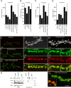
Figure 1.
Figure 1.
FasII signaling through APPL stimulates NMJ growth, and both proteins form a complex in vivo. A-D, Histograms showing the number of boutons at muscles 6 and 7 (abdominal segment 3) of third instar larvae with different levels of FasII and APPL. The number of samples quantified is as follows: wild type, n = 95; fasIIe76/+, n = 12; [FasII]-pre, n = 43; [FasII]-post, n = 17; [FasII]-pre-post (using the Gal4 drivers C380 and BG487), n = 13; [FasII]-pre-post# (using the Gal4 drivers C164 and C57), n = 10; Appld, n = 49; [APPL]-pre (using Gal4 driver C380), n = 14; [APPL]-pre* (using the Gal4 driver C164), n = 85; Appld, fasIIe76/Appld, +, n = 24; Appld, [FasII]-pre-post (using the Gal4 drivers C164 and C57), n = 10; [GPI-FasII]-pre-post (using the Gal4 drivers C380 and BG487), n = 6; [FasII, APPL]-pre-post# (using the Gal4 drivers C380 and BG487), n = 27; [FasII, APPL]-pre, n = 25; [FasII, APPLΔC]-pre, n = 10; fasIIe76, [APPL]-pre, n = 21.Bouton numbers are mean values ± SEM. *p < 0.05; ***p < 0.0001. Low- (E-G) and high- (I-Q) magnification views of third instar larval NMJs at muscles 6 and 7 stained with anti-HRP (E-G, I, J, L-N, P, Q), the synaptic vesicle marker anti-synapsin (K, L, O, P), and anti-HRP and anti-FasII (Q, inset). E, I-L, Wild type. F, M-P, Larvae overexpressing only FasII in both presynaptic and postsynaptic cells. G, Q, Larvae overexpressing both APPL and FasII in both presynaptic and postsynaptic cells. I, M, Stereoscopic images showing budding boutons in wild type (I) and [FasII]-pre-post (M). Note the dramatic increase in budding boutons in larvae overexpressing FasII either alone or in combination with APPL. Scale bars: (in Q) E-G, 50 μm; I-Q, 8 μm. H, FasII and APPL coimmunoprecipitate from body-wall muscle extracts. Extracts were immunoprecipitated with anti-APPL antibodies, and immunoblots were sequentially probed using anti-APPL, anti-FasII, antitubulin (Tub), and anti-spectrin (Spec). Input lanes correspond to 10% of the extract used for immunoprecipitation. Control lanes correspond to extracts in which antibody was omitted during immunoprecipitation. Note that anti-APPL immunoprecipitates FasII and that this interaction is specific because no FasII band could be detected in Appld. Molecular weights are indicated in kilodaltons to the right of each blot. Error bars represent SEM.
Figure 2.
Figure 2.
Electrophysiological analysis of APPL and FasII genetic variants. A, Representative traces of evoked EJPs, in which each trace is the average of 400 EJPs, in wild-type; Appld; fasIIe76/+; Appld, fasIIe76/+; [FasII]-pre-post; Appld, [FasII]-pre-post; and [APPL]-pre. B, Representative mEJP traces of the same genotypes shown in A. C-F, Histograms showing mean ± SEM of evoked EJP amplitude (C), mEJP amplitude (D), mEJP frequency (E), and quantal content (F). A single asterisk indicates significance in relation to wild type; triple carets or triple asterisks indicate p < 0.0001; double carets or double asterisks indicate p < 0.05. Calibration: A, 10 mV, 20 ms; B, 7 mV, 100 ms. Error bars represent SEM.
Figure 3.
Figure 3.
Gross asymmetric changes in FasII levels interfere with APPL-mediated synaptic growth. A-C, Low-magnification views of third instar larval NMJs at muscles 6 and 7 stained with anti-HRP antibodies in wild type (A), a mutant presynaptically overexpressing both APPL and FasII (B), and a larva expressing FasII and APPLΔC presynaptically (C). B, Note the dramatically enlarged boutons in the [FasII, APPL]-pre (arrows) and the long stretches of neuritic processes lacking varicosities (arrowheads). D-R, High-magnification single confocal slices through a bouton in wild type (D, E, I, M, P), [FasII, APPL]-pre (H, L, F, J, N, Q), and [FasII, APPLΔC]-pre (G, K, O, R). Note the internal inclusions and abnormal APPL accumulations in mutants expressing both FasII and APPL and the lack of inclusions and APPL accumulations in samples in which a form of APPL lacking the cytoplasmic domain (ΔC) is expressed. L, Image at higher magnification shows anti-APPL accumulations (arrows) in a [FasII, APPL]-pre bouton. M-R, High-magnification single confocal sections showing anti-FasII (M-O) and anti-HRP and anti-FasII (P-R) in wild type (M, P), [FasII, APPL]-pre (N, Q), and [FasII, APPLΔC]-pre (O, R). Note that the internal FasII-positive inclusions in boutons expressing both APPL and FasII only presynaptically is suppressed when the APPL cytoplasmic domain is absent, demonstrating the specificity of the interaction between APPL and FasII. Scale bars: (in A) A-C, 50 μm; I, 3 μm; D-K, M-R, 5 μm.
Figure 4.
Figure 4.
Microtubule tangles and abnormal APPL deposits are observed at NMJs of [FasII, APPL]-pre larvae. A-C, E-J, NMJs from wild type (A-C) and larvae overexpressing presynaptic FasII and APPL (E-J) showing anti-Futsch (B, C, F, G, I, J), anti-HRP (A, C, E, G, H, J), and merged panels (C, G, J). D, K, Anti-HRP and anti-tubulin in wild type (D) and [FasII, APPL]-pre (K). Note the disorganized appearance of Futsch and microtubules in mutant NMJs (K, arrow). Scale bar: (in K) A-C, E-J, 10 μm; D, K, 6.5 μm.
Figure 5.
Figure 5.
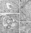
Electron microscopy of abnormal bouton structure in [FasII, APPL]-pre mutants. A-D, Midline cross-sections through wild type (A) and [FasII, APPL]-pre (B-D) boutons. Note the presence of three groups of abnormal concentric rings (C, asterisks) of internal membranes (C, arrowheads) in the mutant, the numerous presynaptic coated vesicles (B, arrowheads), and the large number of postsynaptic vesicle-like structures (D, arrowheads). b, Bouton; SSR, subsynaptic reticulum; arrows point to presynaptic densities. Scale bar: (in C) A, C, 0.8 μm; B, D, 0.4 μm.
Figure 6.
Figure 6.
dX11 and APPL are found in a complex at the NMJ, and changes in dX11 levels mimic changes in APPL levels. A-C, G-I, L-N, Single confocal slices of NMJs stained with anti-dX11 (A, G, L), anti-HRP (B, H, M), and merged panels (C, I, N). A-O, Specimens are wild type (A-E), larvae overexpressing dX11 presynaptically ([dX11]-pre; F-J), and larvae expressing a dX11 variant missing the PTB domain ([ΔPTB]-pre; K-O). D-F, J-K, O, High-magnification views of boutons from the above genotypes, showing the localization of dX11 in each case. Note that, in wild type and [dX11]-pre, dX11 staining is enriched at the presynaptic membrane (E, J), but only in [dX11]-pre does the protein accumulate at sites of budding (F, arrow). K, In contrast, in dX11ΔPTB, dX11 immunoreactivity is observed within the bouton cytoplasm. I, F, Arrows point to buds accumulating dX11. H, Arrowheads point to buds. Scale bar: (in O) A-C, G-I, L-N, 12 μm; D-F, J-K, O, 3 μm.
Figure 7.
Figure 7.
FasII stimulates NMJ expansion by a mechanism that requires both APPL and dX11, and dX11 exists in the APPL-FasII complex. A, Antibodies against dX11 were used to immunoprecipitate dX11 from body-wall muscles of wild type, APPL gain- and loss-of-function mutants, and a strain expressing APPL but lacking the dX11-interacting domain (APPLΔCi). The blot was sequentially blotted with antibodies against dX11, APPL, FasII, spectrin (Spec), and tubulin (Tub). B, Anti-dX11 antibody was used to immunoprecipitate dX11 from body-wall muscle extracts of larvae expressing a full-length dX11 and larvae expressing dX11 lacking the APPL interaction domain (dX11ΔPTB) and sequentially probed with antibodies against dX11, APPL, and tubulin. C, Antibodies against APPL were used to immunoprecipitate APPL from Drosophila S2 cells expressing FasII and/or dX11. In some cases, cells were also treated with dX11 dsRNA. Blots were sequentially probed with antibodies against APPL, FasII, and dX11. D, Western blots of body-wall muscle extracts from wild-type and dX11P/Df larvae probed with antibodies against dX11, APPL, FasII, and tubulin. E, The histogram shows the total number of boutons at muscles 6 and 7 (abdominal segment 3) of third instar wild type, FasII, APPL, and dX11 genetic variants. F, Bouton numbers have been normalized by muscle surface area. The number of samples quantified in E is as follows: wild type, n = 95; Appld, n = 49; [FasII]-pre-post, n = 13, [dX11]-pre, n = 8; Appld, [dX11]-pre, n = 13; [ΔPTB], n = 14; [ΔPTB]#, n = 10; [ΔPTB]-pre-post, n = 8. The number of samples quantified for F is as follows: wild type, n = 30; Appld, n = 23; [FasII]-pre-post, n = 13, [dX11]-pre, n = 14; Appld, [dX11]-pre, n = 13; [ΔPTB], n = 14; [ΔPTB]-pre-post, n = 8; dX11P/Df = 16. *p < 0.05; ***p < 0.0001. Error bars represent SEM.
Figure 8.
Figure 8.
The localization of APPL at synaptic boutons is disrupted in dX11 mutants and a model of likely interactions between FasII, dX11, and APPL during bouton budding. A-D, F-U, Single confocal slices of representative boutons stained with anti-APPL (A, F, J, N, R), anti-APPL and anti-HRP (B, G, K, O, S), anti-FasII (C, H, L, P, T), and anti-FasII and anti-HRP (D, I, M, Q, U), in wild type (A-D), dX11P/Df (F-K), Appld (L, M), [dX11]-pre (N-Q), and [dX11ΔPTB]-pre (R-U). Note that APPL is found at very low levels at the NMJ of wild type. In dX11 mutants and in boutons from larvae overexpressing dX11ΔPTB, there are large internal accumulations of APPL. FasII staining changes little, if any, among the various dX11 specimens. Scale bar, 3.5 μm. E, Diagram depicting likely interactions between FasII, APPL, and dX11 during bouton budding (see Discussion). Eii, Homophilic binding between FasII molecules at opposing sides of the synapse triggers the activation of APPL, an event that may involve its phosphorylation by the tyrosine kinase Abl. Eiii, APPL phosphorylation allows binding to the PTB domain of dX11, which brings or facilitates the insertion of APPL into the presynaptic membrane. dX11 also binds members of the exocytic machinery, thus bringing these components to the budding bouton. In addition, binding of APPL to FasII activates Go protein, which modulates the microtubule cytoskeleton required for bud extension.

References

    1. Ando K, Iijima KI, Elliott JI, Kirino Y, Suzuki T (2001) Phosphorylation-dependent regulation of the interaction of amyloid precursor protein with Fe65 affects the production of beta-amyloid. J Biol Chem 276: 40353-40361. - PubMed
    1. Aplin AE, Gibb GM, Jacobsen JS, Gallo JM, Anderton BH (1996) In vitro phosphorylation of the cytoplasmic domain of the amyloid precursor protein by glycogen synthase kinase-3beta. J Neurochem 67: 699-707. - PubMed
    1. Bailey CH, Chen M, Keller F, Kandel ER (1992) Serotonin-mediated endocytosis of apCAM: an early step of learning-related synaptic growth in Aplysia Science 256: 645-649. - PubMed
    1. Bailey CH, Kaang BK, Chen M, Martin KC, Lim CS, Casadio A, Kandel ER (1997) Mutation in the phosphorylation sites of MAP kinase blocks learning-related internalization of apCAM in Aplysia sensory neurons. Neuron 18: 913-924. - PubMed
    1. Beggs HE, Baragona SC, Hemperly JJ, Maness PF (1997) NCAM140 interacts with the focal adhesion kinase p125(fak) and the SRC-related tyrosine kinase p59(fyn). J Biol Chem 272: 8310-8319. - PubMed

Publication types

MeSH terms