. 2005 Jun 22;25(25):5956-66.
doi: 10.1523/JNEUROSCI.0880-05.2005.

Long-term potentiation is impaired in middle-aged rats: regional specificity and reversal by adenosine receptor antagonists

Affiliations

Long-term potentiation is impaired in middle-aged rats: regional specificity and reversal by adenosine receptor antagonists

Christopher S Rex et al. J Neurosci. .

Abstract

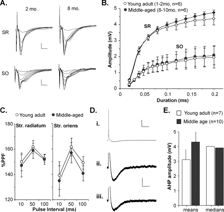
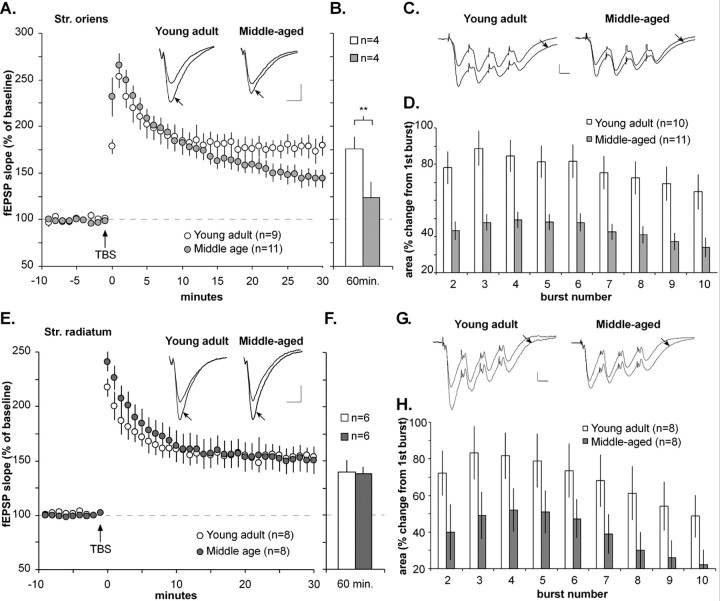
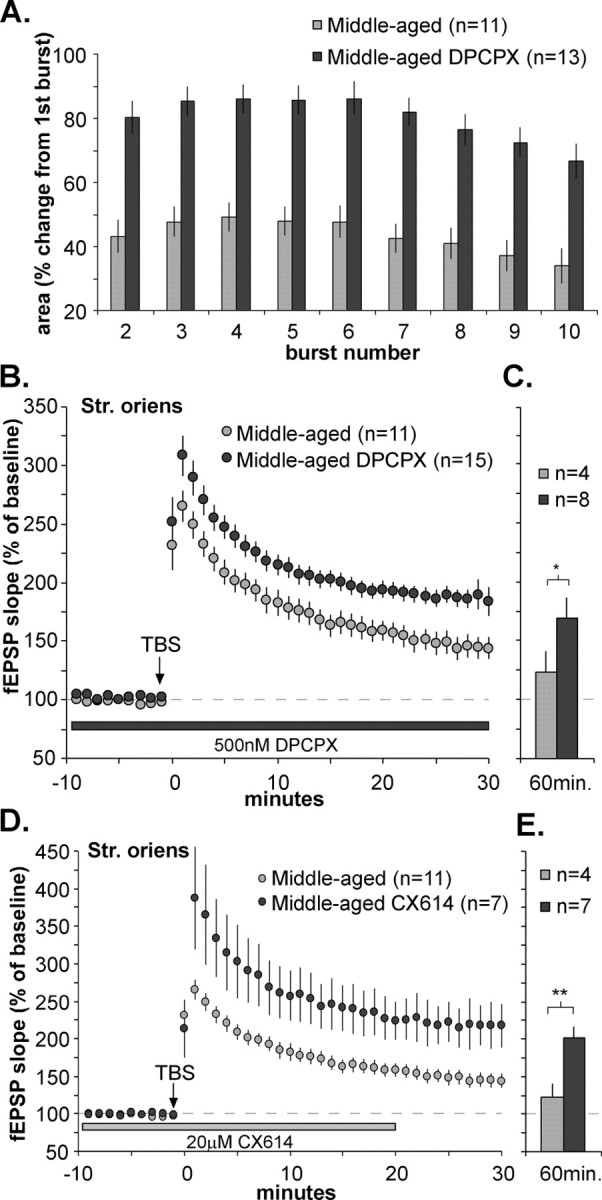
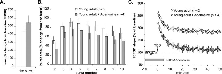
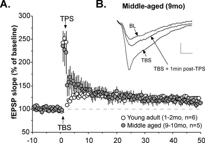
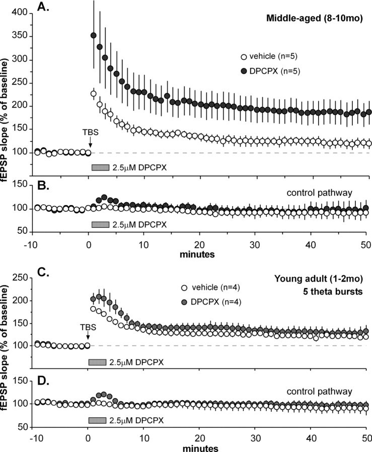
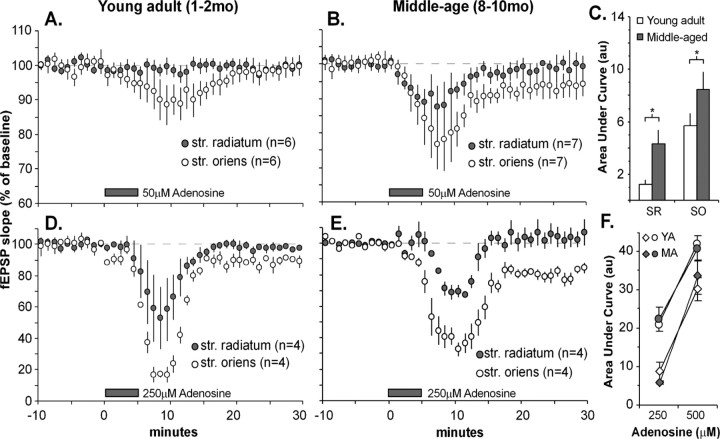
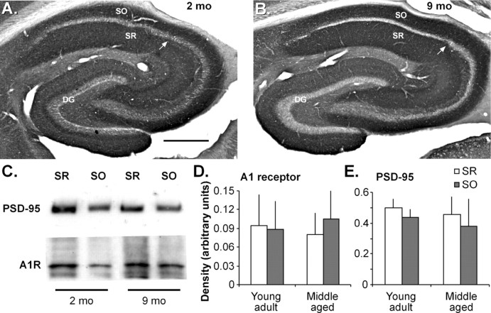
Memory loss in humans begins early in adult life and progresses thereafter. It is not known whether these losses reflect the failure of cellular processes that encode memory or disturbances in events that retrieve it. Here, we report that impairments in hippocampal long-term potentiation (LTP), a form of synaptic plasticity associated with memory, are present by middle age in rats but only in select portions of pyramidal cell dendritic trees. Specifically, LTP induced with theta-burst stimulation in basal dendrites of hippocampal field CA1 decayed rapidly in slices prepared from 7- to 10-month-old rats but not in slices from young adults. There were no evident age-related differences in LTP in the apical dendrites. Both the adenosine A1 receptor antagonist 8-cyclopentyl-1,3-dipropylxanthine and a positive AMPA receptor modulator (ampakine) offset age-related LTP deficits. Adenosine produced greater depression of synaptic responses in middle-aged versus young adult slices and in basal versus apical dendrites. These results were not associated with variations in A1 receptor densities and may instead reflect regional and age-related differences in adenosine clearance. Pertinent to this, brief applications of A1 receptor antagonists immediately after theta stimulation fully restored LTP in middle-aged rats. We hypothesize that the build-up of extracellular adenosine during theta activity persists into the postinduction period in the basal dendrites of middle-aged slices and thereby activates the A1 receptor-dependent LTP reversal effect. Regardless of the underlying mechanism, the present results provide a candidate explanation for memory losses during normal aging and indicate that, with regard to plasticity, different segments of pyramidal neurons age at different rates.


Figures

Figure 1.
Figure 1.
Young adult and middle-aged Schaffer-commissural responses are comparable by several measures. A, Traces from representative input-output experiments comparing fEPSP responses within the stratum radiatum (SR) and stratum oriens (SO) between slices of 2- and 8-month-old rats. Calibration: 0.5 mV, 10 ms. B, Input-output curves for the apical (SR) and basal (SO) branches of the Schaffer-commissural projections in young adult and middle-aged slices. The shape of the curves differs for apical versus basal locations, but there are no evident age-related differences. C, Paired-pulse facilitation recorded from SR and SO at 10, 50, and 100 ms pulse intervals showed no differences between slices from young adult and middle-aged rats. D, Representative whole-cell clamp recordings from slices of 9-month-old rats. Stimulation of Schaffer-commissural axons in the stratum oriens elicited EPSCs that are fully developed, shown at low (Di) and high (Dii, Diii) magnification. Calibration: Di, 20 mV, 0.5 s; Dii, Diii, 2.5 mV, 0.5 s. E, Comparison of mean (left) AHP amplitudes, measured from EPSCs of young adult and middle-aged slices, reveals no significant age-related differences (p > 0.2; two-tailed t test). Median scores (right) are nearly identical.
Figure 2.
Figure 2.
LTP fails to stabilize in basal, but not apical, dendrites from 7- to 10-month-old rats. A, Plot of group data for the Schaffer-commissural fEPSP slope within the basal dendritic field of hippocampal slices from young adult (1-2 months of age) and middle-aged (8-10 months of age) rats before and after LTP induction. TBS was applied to induce LTP after a 10 min baseline at the time indicated by an arrow. As shown, middle-aged slices showed no deficits in the magnitude of LTP within the first 10 min after a train of 10 theta bursts but deviated from the young adult pattern by continuing to decay thereafter. Inset, Representative traces recorded from the str. oriens during the baseline recording period and 30 min after TBS (arrow). Calibration: 0.5 mV, 10 ms. B, Slope of the fEPSP in a subset of young adult and middle-aged slices 60 min after TBS; values indicate that LTP remained stable in young adult slices but continued to decline in the middle-aged group (**p < 0.01; two-tailed t test). C, Burst responses recorded during TBS. Averaged responses (from a subset of 4 rats) from burst 1 and burst 4 (arrow) are superimposed. Facilitation from burst 1 to burst 4 is notably smaller in slices from middle-aged rats compared with young adults. Calibration: 0.5 mV, 10 ms. D, Comparison of the size of theta-burst responses within a theta train in young adult and middle-aged rats: the plot shows the mean change in the area of responses 2-10, relative to the first theta-burst response; the middle-aged slices had much smaller within-train facilitation (average area of bursts 2-10, 43 ± 4.7%) compared with slices from young adults (77 ± 8.6%). E, Plot of group data showing the Schaffer collateral/commissural fEPSP slope within the str. radiatum of young adult and middle-aged slices before and after TBS. As is apparent, the fEPSP slopes in both groups were comparable across the 30 min testing period after TBS. Inset, Representative fEPSPs recorded from the str. radiatum in slices from young adult and middle-aged rats during the baseline recording period and 30 min after TBS (arrow). Calibration: 0.5 mV, 10 ms. F, Potentiation in the apical dendrites of young adult and middle-aged slices was equivalent 60 min after TBS. G, Averaged responses (from a subset of 4 rats) during burst 1 and burst 4 (arrow) are overlaid for young adult and middle-aged rats. Note that the middle-aged group exhibited reduced facilitation in the apical dendrites, as was the case for the basal dendrites. Calibration bars: 0.5 mV, 10 ms. H, Comparison of the size of theta-burst responses within a TBS train in young and middle-aged rats: the plot shows the mean change in the area of burst responses 2-10 relative to the size of the first theta-burst response. Slices prepared from middle-aged rats had significantly smaller within-train facilitation in the apical dendrites than did those from young adults.
Figure 3.
Figure 3.
The adenosine A1 antagonist DPCPX and an AMPA receptor-positive modulator rescue LTP stabilization in middle-aged basal dendrites. The effects of TBS on Schaffer-commissural fEPSPs within the stratum oriens were evaluated in slices from middle-aged rats with and without 500 nm DPCPX treatment. A, Comparison of the composite area of burst responses within a theta train in slices from middle-aged rats with and without DPCPX. The graph summarizes the mean percentage change in the area of burst responses 2-10 relative to the size of the first burst response. DPCPX significantly increased theta train facilitation, bringing it into the range of facilitation levels seen in young adult tissue (compare with Fig. 2 D). B, Plot of fEPSP slope before and after TBS (values normalized to the mean response during the last 10 min of the baseline period in the presence of DPCPX). As shown, DPCPX treatment enhanced the magnitude of LTP in middle-aged slices at all time points. C, DPCPX-treated slices from middle-aged animals tested for 1 h after TBS showed stable potentiation that was significantly enhanced compared with middle-aged control slices (*p < 0.05; two-tailed t test). D, A positive modulator of AMPA receptors also enhances LTP in the basal dendrites of middle-aged rats. The percentage of LTP induced by a single train of theta bursts was measured in the stratum oriens in the presence and absence of CX614 (20 μm). The graph of fEPSP slopes is expressed as percentage of the mean baseline response during the 10 min preceding TBS in the presence of CX614; slices infused with CX614 had greater LTP during the 30 min after TBS. E, The positive effects of CX614 were maintained throughout the 60 min after TBS. (**p < 0.01 compared with drug-control values; two-tailed t test).
Figure 4.
Figure 4.
Low concentrations of adenosine do not block theta-burst facilitation but do prevent LTP consolidation in the stratum oriens of young adult slices. A, The areas of single theta-burst responses (i.e., the composite potential generated by 4 pulses at 100 Hz) are expressed as a fraction of baseline fEPSP area (during the 5 min preceding TBS). Group values show that the burst area is increased over the fEPSP area to the same degree in adenosine-treated (gray bar) and control (white bar) slices. B, The plot shows the mean change in the size of burst responses 2-10, relative to the size of the first theta-burst response within a theta train; facilitation of burst responses was not substantially affected by infusion of 750 nm adenosine. C, A plot showing the effects of TBS on Schaffer-commissural fEPSP slopes with and without bath adenosine treatment in the basal dendritic field of young adult slices. Adenosine was added to perfusion lines at 750 nm for 40 min before the delivery of TBS and washed out 5 min afterward. As is apparent, exogenous adenosine completely suppressed the stabilization of LTP (2-way repeated-measures ANOVA; p < 0.01).
Figure 5.
Figure 5.
The LTP reversal effect is fully operational in the basal dendrites of middle-aged slices. TBS was applied to Schaffer-commissural projections in the stratum oriens in slices prepared from young adult (1-2 months of age) or middle-aged (9-10 months of age) rats. A, B, A single train of 5 Hz stimulation (TPS) was delivered to the potentiated pathway beginning 60 s after TBS. A, The graph summarizes group results and confirms that potentiation did not recover but instead decayed almost to baseline over the following 30 min. B, As seen in the traces from a representative experiment, the 5 Hz stimulation caused an immediate and pronounced loss of potentiation in middle-aged slices.
Figure 6.
Figure 6.
Adenosine A1 receptor antagonist applied after TBS rescues robust LTP in middle-aged slices. TBS was applied to the Schaffer-commissural system in the str. oriens, immediately after which the A1 receptor antagonist DPCPX or vehicle was infused from an adjacent micropipette controlled via an automated pump. fEPSPs were simultaneously recorded from a second collection of Schaffer-commissural projections (the “control pathway”) to confirm that the drug was working in the DPCPX cases and to ensure that the infusion itself did not disrupt baseline recording in the control (vehicle) cases. A, Slices in which TBS was followed by vehicle infusion (open circles) exhibited an LTP effect that did not stabilize but instead decayed almost to baseline during the postinduction period (baseline indicated by dashed line). In contrast, a 3 min infusion of DPCPX immediately after TBS (filled circles) caused potentiation to stabilize at 83 ± 24.7% above baseline (i.e., values comparable with those found in young adult slices) at 50 min after induction. B, Effects of vehicle or DPCPX infusions on the converging Schaffer-commissural control pathway that did not receive TBS. As shown, slices treated with DPCPX, but not vehicle, exhibited a brief response facilitation, indicating that the A1R antagonist quickly reached the recording sites at effective concentrations and subsequently washed out with little delay. C, Suboptimal stimulation (5 theta bursts) was delivered to the str. oriens of young adult slices, followed immediately by a 3 min infusion of DPCPX (filled circles) or vehicle (open circles). By 50 min after stimulation, DPCPX-treated slices did not show greater potentiation than vehicle-treated slices (p > 0.5; 2-way repeated-measures ANOVA for minutes 40-50), indicating that blocking A1 receptors after stimulation does not increase maximum LTP. D, Effects of vehicle or DPCPX infusions on the nonstimulated control pathway.
Figure 7.
Figure 7.
Region- and age-related differences in adenosine effects on synaptic responses. High concentrations of adenosine were added to perfusion lines for 5 min and then washed out for 25 min. fEPSPs generated by stimulation of the apical and basal branches of the Schaffer-collateral/commissural projections were recorded simultaneously in the str. radiatum and str. oriens, respectively. A, B, Adenosine at 50 μm caused a rapid reduction of fEPSP slope that was greater in the str. oriens than in the str. radiatum in both young adult (A) and middle-aged (B) slices. C, Quantification of the area under the curve (i.e., the deviation from baseline as plotted in A and B and expressed in arbitrary units) from the time of initial infusion of 50 μm adenosine to 15 min after drug washout. Responses for the str. radiatum (SR) and str. oriens (SO) show age-related differences in drug sensitivity in both strata (p > 0.05; two-tailed t test). D, E, High concentrations of adenosine (250 μm) markedly depressed Schaffer-collateral/commissural fEPSPs with the same pattern of regional differences as that seen at the lower concentration. Note that washout was not completely effective in the str. oriens of the middle-aged slices. F, Dose-response relationships using area under the curve as a response measure. Differences were not noted between young adult (YA; open symbols) and middle-aged (MA; filled symbols) slices with high-concentration adenosine infusions (250 and 500 μm), but striking differences between SO (circles) and SR (diamonds) were observed for both age groups.
Figure 8.
Figure 8.
Regional adenosine A1 receptor levels in the hippocampus of rats of different ages. A, B, Low-magnification photomicrographs of immunostaining for the A1 receptor in sections through the hippocampus of 2-month-old (A) and 9-month-old (B) rats shows comparably dense immunostaining in the CA1 str. oriens (SO) and str. radiatum (SR). The arrow indicates the approximate CA1/CA2 boundary. Scale bar: (in A) A, B, 500 μm. C, Representative Western blot shows levels of PSD-95 (top) and A1 receptor (A1R; bottom) immunoreactivities in homogenates from the CA1 str. radiatum (SR) and str. oriens (SO) from young adult (1-2 months of age) and middle-aged (8-10 months of age) rats (10 μg of protein per lane; immunoreactive bands were 34 and 89 kDa, respectively). D, E, Graphs show levels of A1 receptor (D) and PSD-95 (E) immunoreactivities assessed by densitometric analysis of Western blot ECL films (n = 6 rats/age).

References

    1. Abraham WC, Huggett A (1997) Induction and reversal of long-term potentiation by repeated high-frequency stimulation in rat hippocampal slices. Hippocampus 7: 137-145. - PubMed
    1. Applegate MD, Landfield PW (1988) Synaptic vesicle redistribution during hippocampal frequency potentiation and depression in young and aged rats. J Neurosci 8: 1096-1111. - PMC - PubMed
    1. Arai A, Lynch G (1992) Factors regulating the magnitude of long-term potentiation induced by theta pattern stimulation. Brain Res 598: 173-184. - PubMed
    1. Arai A, Larson J, Lynch G (1990a) Anoxia reveals a vulnerable period in the development of long-term potentiation. Brain Res 511: 353-357. - PubMed
    1. Arai A, Kessler M, Lynch G (1990b) The effects of adenosine on the development of long-term potentiation. Neurosci Lett 119: 41-44. - PubMed

Publication types

Substances