. 2005 Jun 22;25(25):6016-24.
doi: 10.1523/JNEUROSCI.0692-05.2005.

Intravesicular localization and exocytosis of alpha-synuclein and its aggregates

Affiliations

Intravesicular localization and exocytosis of alpha-synuclein and its aggregates

He-Jin Lee et al. J Neurosci. .

Abstract

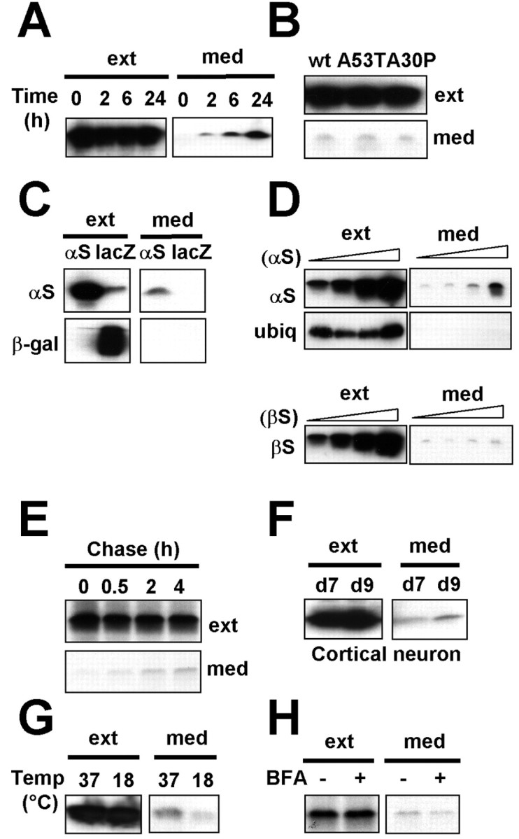
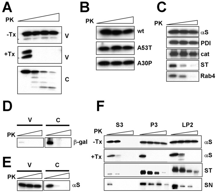
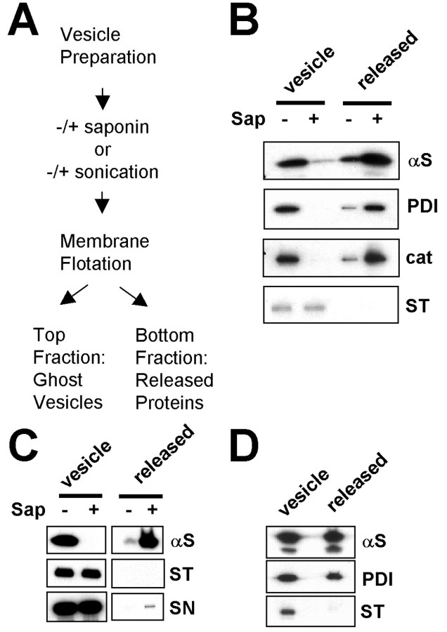
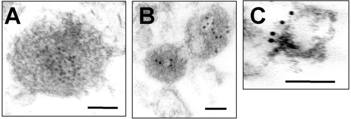
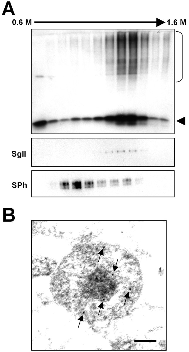
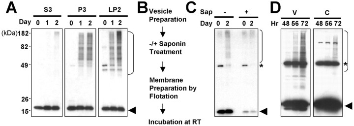
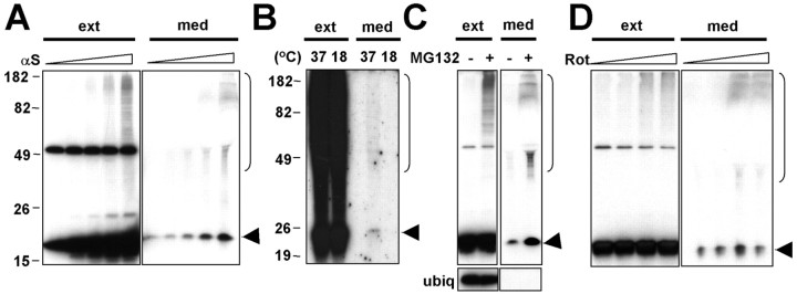
Alpha-synuclein (alpha-syn), particularly in its aggregated forms, is implicated in the pathogenesis of Parkinson's disease and other related neurological disorders. However, the normal biology of alpha-syn and how it relates to the aggregation of the protein are not clearly understood. Because of the lack of the signal sequence and its predominant localization in the cytosol, alpha-syn is generally considered exclusively an intracellular protein. Contrary to this assumption, here, we show that a small percentage of newly synthesized alpha-syn is rapidly secreted from cells via unconventional, endoplasmic reticulum/Golgi-independent exocytosis. Consistent with this finding, we also demonstrate that a portion of cellular alpha-syn is present in the lumen of vesicles. Importantly, the intravesicular alpha-syn is more prone to aggregation than the cytosolic protein, and aggregated forms of alpha-syn are also secreted from cells. Furthermore, secretion of both monomeric and aggregated alpha-syn is elevated in response to proteasomal and mitochondrial dysfunction, cellular defects that are associated with Parkinson's pathogenesis. Thus, intravesicular localization and secretion are part of normal life cycle of alpha-syn and might also contribute to pathological function of this protein.


Figures

Figure 1.
Figure 1.
α-Syn is secreted from cells via exocytosis. A, Release of α-syn from differentiated SH-SY5Y cells. The conditioned medium (med) and the whole-cell extracts (ext) were collected at the indicated times. B, Release of α-syn mutants from differentiated SH-SY5Y cells. wt, Wild type. C, Overexpression of β-galactosidase does not cause its release. The conditioned medium and the cell extracts were collected from differentiated SH-SY5Y cells overexpressing either α-synorlacZ (β-gal). D, Secretion of α-syn correlates with its expression levels. Different doses of recombinant adenoviruses, adeno/α-syn and adeno/β-syn (1.25, 5, 20, 80 m.o.i.), were expressed in differentiated SH-SY5Y cells, and the levels of intracellular (ext) and released (med) proteins were analyzed. Released proteins were collected for 3 h. Note that ubiquitin (ubiq) was not released from the cells overexpressing α-syn. E, Pulse-chase experiment of α-syn secretion. SH-SY5Y cells expressing α-syn-MycHis were metabolically labeled with 35S-met/cys for 30 min and chased for indicated periods, and α-syn-MycHis was purified from the conditioned medium and the cell extract using Ni-NTA agarose beads. F, Secretion of endogenous α-syn from primary cortical neurons. The medium and the cell extracts were collected at day 7 (d7) or day 9 (d9) after 48 h of incubation in fresh medium. G, α-Syn secretion is blocked by low-temperature incubation. SH-SY5Y cells expressing α-syn were incubated at either 37 or 18°C for 3 h. H, Secretion of α-syn is insensitive to BFA. SH-SY5Y cells expressing α-syn-MycHis were pretreated with BFA for 2 h and metabolically labeled with 35S-met/cys for 30 min and chased for 2 h in the presence of BFA. αS, α-Syn; βS, β-syn.
Figure 2.
Figure 2.
Vesicular α-syn is not accessible to protease digestion. A, PK resistance of vesicular α-syn. V and C prepared from differentiated SH-SY5Y cells were incubated with different concentrations of PK. Note that detergent-mediated solubilization of membrane (1% Triton X-100) eliminates PK resistance of vesicular α-syn. B, PK resistance of mutant α-syn in vesicles. Microsomal vesicles were prepared from differentiated SH-SY5Y cells overexpressing wild type (wt) or mutants and incubated with different concentrations of PK (0, 0.1 and 1 μg/ml). C, PK digestion of other vesicular proteins. Microsomal vesicles from SH-SY5Y cells were incubated with PK. Concentrations of PK: 0, 0.1 and 1 μg/ml. D, PK sensitivity of β-galactosidase (β-gal). V and C from SH-SY5Y cells overexpressing β-galactosidase were treated with 0, 0.1, and 1 μg/ml PK. E, PK sensitivity of neuronal endogenous α-syn. Vesicular and cytosolic fractions were isolated from rat cortical neurons and treated with PK. F, PK sensitivity of brain α-syn. Microsomal vesicles (P3), crude synaptic vesicles (LP2), and cytosol (S3) were prepared from rat brain homogenate and subjected to PK digestion. +Tx, With 1% Triton X-100; -Tx, without Triton X-100; αS, α-syn; cat, catalase; ST, synaptotagmin 1; SN, synapsin 1.
Figure 3.
Figure 3.
Vesicular α-syn is released from vesicles after membrane permeabilization. A, Diagram of the lumenal protein release experiment. Vesicles were prepared from differentiated SH-SY5Y cells overexpressing α-syn or rat brain tissues and were permeabilized with saponin treatment or sonication. Saponin treatment and sonication produced ghost vesicles without lumenal components. The ghost vesicles and released proteins were separated with the flotation centrifugation. B, Lumenal protein release from the SH-SY5Y vesicles after the saponin-mediated permeabilization. C, Lumenal protein release from the rat brain P3 vesicles after the saponin-mediated permeabilization. D, Protein release from the SH-SY5Y vesicles after the sonication-mediated permeabilization. Sap, Saponin; αS, α-syn; cat, catalase; ST, synaptotagmin 1; SN, synapsin 1.
Figure 4.
Figure 4.
Immunoelectron microscopy of the brain vesicles and neuroblastoma vesicles. Rat brain P3 vesicles (A), SH-SY5Y vesicles (B), and rat brain crude synaptic vesicles (C) were prepared by the flotation centrifugation and examined after immunogold labeling for α-syn (A, B) and synapsin 1 (C). Note that 10 nm of gold particles label α-syn in the lumen of electron-dense vesicles and synapsin-1 on the surface of vesicles. Scale bars, 100 nm.
Figure 5.
Figure 5.
α-Syn in LDCVs. A, Subcellular fractionation of LDCVs. Cell extract from differentiated SH-SY5Y cells overexpressing α-syn was subjected to a linear sucrose gradient centrifugation, and fractions were collected from the top. High-molecular-weight aggregates are marked with a bracket on the right, and monomeric α-syn is marked with an arrowhead. SgII, Secretogranin II; SPh, synaptophysin. B, Immunoelectron microscopy of LDCV fractions (fractions 8 and 9). Arrows indicate 10 nm of gold particles labeling α-syn. Scale bar, 100 nm.
Figure 6.
Figure 6.
Aggregation of the vesicular α-syn. A, Rate of α-syn aggregation in vesicles and cytosol. Rat brain fractions were incubated at room temperature for 1 or 2 d. High-molecular-weight aggregates are marked with a bracket on the right, and monomeric α-syn is marked with an arrowhead. B, C, Lumenal α-syn is responsible for vesicular α-syn aggregation. Vesicles were prepared from SH-SY5Y cells overexpressing α-syn and treated with either a buffer with (+) or without (-) saponin. Treated vesicles were isolated once again with flotation centrifugation and incubated for 2 d.α-Syn aggregation was analyzed by Western blotting (C). The asterisk indicates an unrelated protein that cross-reacts to Syn-1 antibody (Perrin et al., 2003). D, Time course of α-syn aggregation in V and C in intact cells. Sap, Saponin; RT, room temperature.
Figure 7.
Figure 7.
Secretion of aggregated α-syn. A, Secretion of α-syn (αS) aggregates parallels with intracellular aggregate formation. SH-SY5Y cells were infected with adeno/α-syn at increasing m.o.i. (0, 2.5, 5, 10, 20). Secreted proteins were collected for 16 h. High-molecular-weight aggregates are marked with a bracket on the right, and monomeric α-syn is marked with an arrowhead. The whitening in the Western data of medium (60 kDa area) is attributable to an abundant presence of serum albumin. B, Secretion of α-syn aggregates is blocked by low-temperature incubation. SH-SY5Y cells expressing α-syn-MycHis were metabolically labeled with 35S-met/cys for 2 h, and the fresh medium was added for 7 h and the medium was changed again and chased for another 2 h at either 37 or 18°C to monitor secretion of aggregates. C, Secretion of α-syn in response to MG132. Cells were treated with 10 μm MG132 for 24 h to induce aggregation, and the medium and the cell extract were collected. D, Secretion of α-syn from rotenone (Rot)-treated cells. Cells were treated with rotenone (0, 3, 10, and 30 nm) for 2 d to induce aggregation. ext, Whole-cell extract; med, conditioned medium; ubiq, ubiquitin.

References

    1. Abeliovich A, Schmitz Y, Farinas I, Choi-Lundberg D, Ho WH, Castillo PE, Shinsky N, Verdugo JM, Armanini M, Ryan A, Hynes M, Phillips H, Sulzer D, Rosenthal A (2000) Mice lacking alpha-synuclein display functional deficits in the nigrostriatal dopamine system. Neuron 25: 239-252. - PubMed
    1. Borghi R, Marchese R, Negro A, Marinelli L, Forloni G, Zaccheo D, Abbruzzese G, Tabaton M (2000) Full length alpha-synuclein is present in cerebrospinal fluid from Parkinson's disease and normal subjects. Neurosci Lett 287: 65-67. - PubMed
    1. Bussell Jr R, Eliezer D (2003) A structural and functional role for 11-mer repeats in alpha-synuclein and other exchangeable lipid binding proteins. J Mol Biol 329: 763-778. - PubMed
    1. Cabin DE, Shimazu K, Murphy D, Cole NB, Gottschalk W, McIlwain KL, Orrison B, Chen A, Ellis CE, Paylor R, Lu B, Nussbaum RL (2002) Synaptic vesicle depletion correlates with attenuated synaptic responses to prolonged repetitive stimulation in mice lacking α-synuclein. J Neurosci 22: 8797-8807. - PMC - PubMed
    1. Chandra S, Chen X, Rizo J, Jahn R, Sudhof TC (2003) A broken alpha-helix in folded alpha-synuclein. J Biol Chem 278: 15313-15318. - PubMed

Publication types

LinkOut - more resources