. 2005 Oct 15;106(8):2693-9.
doi: 10.1182/blood-2005-03-1131. Epub 2005 Jun 23.

Dose-dependent effects of the Notch ligand Delta1 on ex vivo differentiation and in vivo marrow repopulating ability of cord blood cells

Affiliations

Dose-dependent effects of the Notch ligand Delta1 on ex vivo differentiation and in vivo marrow repopulating ability of cord blood cells

Colleen Delaney et al. Blood. .

Abstract

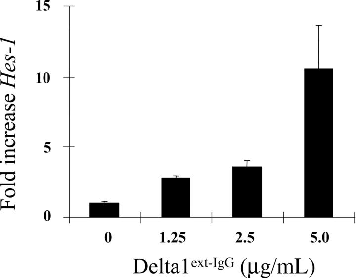
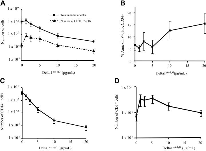
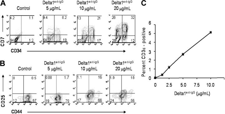
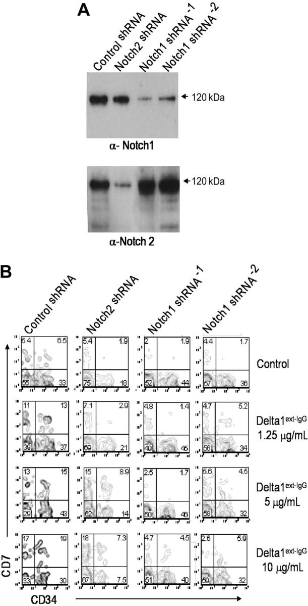
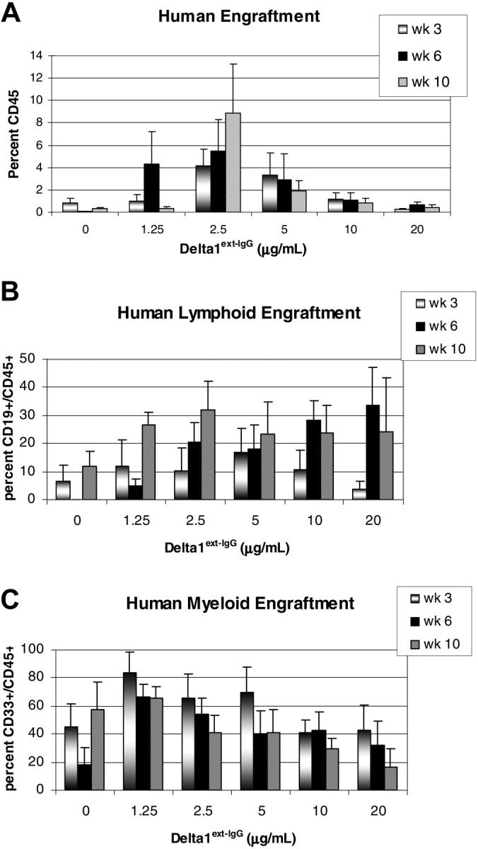
Although significant advances have been made over the last decade with respect to our understanding of stem cell biology, progress has been limited in the development of successful techniques for clinically significant ex vivo expansion of hematopoietic stem and progenitor cells. We here describe the effect of Notch ligand density on induction of Notch signaling and subsequent cell fate of human CD34+CD38- cord blood progenitors. Lower densities of Delta1(ext-IgG) enhanced the generation of CD34+ cells as well as CD14+ and CD7+ cells, consistent with early myeloid and lymphoid differentiation, respectively. However, culture with increased amounts of Delta1(ext-IgG) induced apoptosis of CD34+ precursors resulting in decreased cell numbers, without affecting generation of CD7+ cells. RNA interference studies revealed that the promotion of lymphoid differentiation was primarily mediated by Delta1 activation of Notch1. Furthermore, enhanced generation of NOD/SCID repopulating cells was seen following culture with lower but not higher densities of ligand. These studies indicate critical, quantitative aspects of Notch signaling in affecting hematopoietic precursor cell-fate outcomes and suggest that density of Notch ligands in different organ systems may be an important determinant in regulating cell-fate outcomes. Moreover, these findings contribute to the development of methodology for manipulation of hematopoietic precursors for therapeutic purposes.


Figures

Figure 1.
Figure 1.
Density-dependent activation of endogenous Notch signaling as indicated by quantitative RT-PCR of Hes1 mRNA. CD34+CD38 cord blood precursors were incubated with increasing densities of Delta1ext-IgG and expression of the Notch target gene, Hes1, was measured after 6 hours by quantitative RT-PCR (SYBR-green). Results indicate the fold increase in Hes1 levels normalized to control human IgG at increasing densities of Delta1ext-IgG. Hes1 levels were corrected for mRNA levels based on housekeeping gene expression (R = 0.92; 95% CI, 0.86-0.97; P < .001). Results are representative of 3 experiments ± SEM.
Figure 2.
Figure 2.
Delta1ext-IgG affects the number and apoptosis of CD34+ precursors and the number of myeloid and lymphoid precursors in a dose-dependent manner. CD34+CD38 cord blood precursors were incubated with increasing Delta1ext-IgG densities or human control IgG (designated in the figures as Delta1ext-IgG at 0 μg/mL). (A) Total number of cells (▪), and number of CD34+ cells (▴) generated following 21 days of culture. Results are the mean of 7 independent experiments ± SEM (R2 = 0.97; P = .002 for total cells and .001 for CD34). (B) Maximum percentage of CD34+ cells that are annexin V positive and propidium iodide negative] following 3 to 6 days in culture. Percent apoptosis was measured on days 3, 6, 10, and 14 in 3 independent experiments and maximum apoptosis of the CD34+ cells was seen at 3 and 6 days. Results are the mean of 3 independent experiments ± SEM. (C-D) Total number of CD14+ (R2 = 0.95; P = .004) and CD7+ cells, respectively, generated following 21 days of culture. Results are the mean of 7 independent experiments ± SEM.
Figure 3.
Figure 3.
Dose-dependent induction of lymphoid differentiation. CD34+CD38 cord blood precursors were incubated with increasing Delta1ext-IgG densities or human control IgG and analyzed by FACS at day 21 for CD7 and CD34 expression (A), day 10 for CD25 and CD44 expression (B), and day 14 for intracellular CD3ε expression (C). Control human IgG is designated in the figures as control or Delta1ext-IgG at 0 μg/mL. Data are representative of 3 independent experiments. Numbers in corners of dot blots are the percentage of gated events within that quadrant (R2 = 0.95; P = .004).
Figure 4.
Figure 4.
Notch1 but not Notch2 is primarily responsible for Notch-induced lymphoid differentiation. CD34+CD38 cord blood cells were transduced with either Notch1, Notch2, or control shRNA constructs and cultured in the presence of increasing concentrations of immobilized Delta1ext-IgG. After 4 days, cells were GFP sorted and replaced in culture. After 7 days, cells were harvested to prepare lysates or immunostain for FACS analysis. (A) Lysates were separated with sodium dodecyl sulfate–polyacrylamide gel electrophoresis, transferred to nitrocellulose, and immunoblotted with the α-Notch1 monoclonal antibody, MN-1, stripped, and reimmunoblotted with the anti-Notch2 monoclonal antibody, BHN6. (B) Cells were assessed for CD7 and CD34 expression. Numbers in corners are the percentage of gated events within that quadrant.
Figure 5.
Figure 5.
Human engraftment in NOD/SCID mice of Delta1ext-IgG cultured cells. CD34+CD38 cord blood precursors were cultured for 3 weeks with increasing Delta1ext-IgG densities or human control IgG (designated in the figures as Delta1ext-IgG at 0 μg/mL) and transplanted into sublethally irradiated NOD/SCID mice. (A) Percent overall human engraftment (CD45+), (B) percent human lymphoid engraftment, and (C) percent human myeloid engraftment were assessed in bone marrow aspirates from mice at 3, 6, and 10 weeks. Data are the results of 5 independent experiments, with the mean ± SEM.

Similar articles

Cited by

References

    1. Izon DJ, Punt JA, Pear WS. Deciphering the role of Notch signaling in lymphopoiesis. Curr Opin Immunol. 2002;14: 192-199. - PubMed
    1. Allman D, Punt JA, Izon DJ, Aster JC, Pear WS. An invitation to T and more: notch signaling in lymphopoiesis. Cell. 2002;109(suppl): S1–S11. - PubMed
    1. Robson-MacDonald H, Wilson A, Radtke F. Notch1 and T-cell development: insights from conditional knockout mice. Trends Immunol. 2001;22: 155-160. - PubMed
    1. Jones P, May G, Healy L, et al. Stromal expression of Jagged 1 promotes colony formation by fetal hematopoietic progenitor cells. Blood. 1998;92: 1505-1511. - PubMed
    1. Ohishi K, Varnum-Finney B, Bernstein ID. Delta-1 enhances marrow and thymus repopulating ability of human CD34+CD38– cord blood cells. J Clin Invest. 2002;110: 1165-1174. - PMC - PubMed

Publication types