. 2005 Jul 18:354:132-9.
doi: 10.1016/j.gene.2005.03.028.

Mitochondria-to-nucleus stress signaling in mammalian cells: nature of nuclear gene targets, transcription regulation, and induced resistance to apoptosis

Affiliations

Mitochondria-to-nucleus stress signaling in mammalian cells: nature of nuclear gene targets, transcription regulation, and induced resistance to apoptosis

Gopa Biswas et al. Gene. .

Abstract

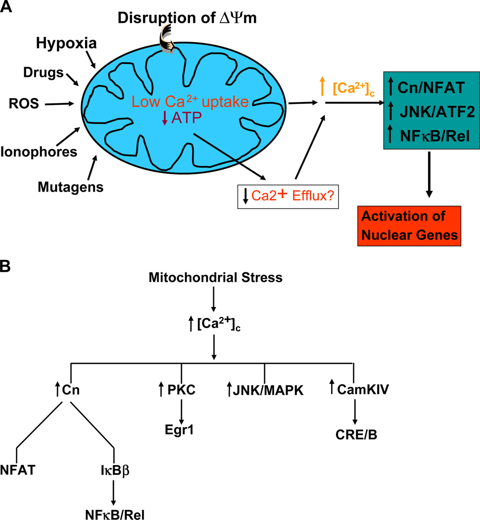
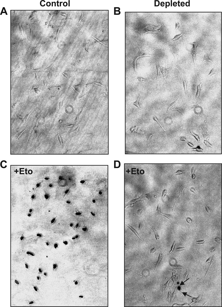
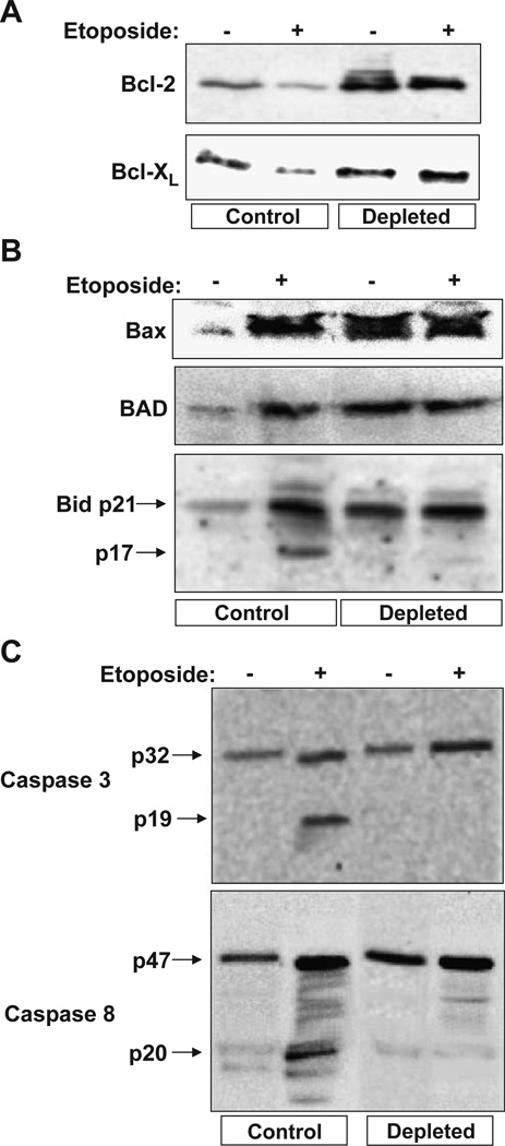
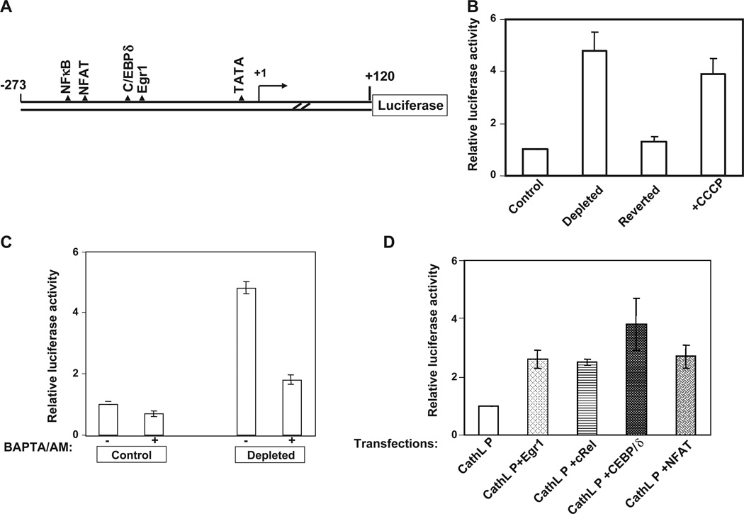
Depletion of mitochondrial DNA (mtDNA) or treatment with mitochondrial poison CCCP initiates mitochondrial stress signaling, which operates through altered Ca2+ homeostasis. In C2C12 rhabdomyoblasts and A549 human lung carcinoma cells mitochondrial stress signaling activates calcineurin and a number of Ca2+ responsive factors including ATF, NFAT, CEBP/delta and CREB. Additionally, PKC and MAP kinase are also activated. A number of nuclear gene targets including those involved in Ca2+ storage/release (RyR1, calreticulin, calsequestrin), glucose metabolism (hexokinase, pyruvate kinase, Glut4), oncogenesis (TGFbeta1, cathepsin L, IGFR1, melanoma antigen) and apoptosis (Bcl-2, Bid, Bad, p53) are upregulated. Mitochondrial stress in both C2C12 myoblasts and A549 cells induced morphological changes and invasive phenotypes. These cells also showed markedly increased resistance to etoposide-induced apoptosis that is a hallmark of highly invasive tumors. Our results describe a new mechanism of altered nuclear gene expression and phenotypic changes triggered by mitochondrial dysfunction and mtDNA damage.


Figures

Fig. 1
Fig. 1
Mitochondrial retrograde signaling in mammalian cells through altered Ca2+ levels and activation of Ca2+ responsive factors. (A) A proposed model for mitochondrial stress signaling caused by disruption of ΔΨm either by mtDNA depletion, or treatment with drugs/chemicals that affect mitochondrial function. An upward arrow indicates a net increase in concentration (such as free Ca2+), activation of factors or upregulation of transcription. A downward arrow indicates inactivation of factors or lower activity. (B) List of transcription factors and signaling proteins known to be activated by different forms of stress induced retrograde signaling.
Fig. 2
Fig. 2
Resistance to etoposide-mediated apoptosis in mtDNA-depleted C2C12 cells. Control (A and C), and mtDNA-depleted (B and D) cells were subjected to apoptosis assay before (A and B) or after treatment with 25 µM etoposide for 4 h to induce apoptosis. The extent of nuclear DNA breaks was measured by TUNEL assay with anti Digoxigenin peroxidase ApopTag Peroxidase system as described in the Materials and methods section.
Fig. 3
Fig. 3
Steady-state levels of pro- and anti-apoptotic proteins and caspases in etoposide-treated control and mtDNA-depleted C2C12 cells. Cells treated with (25 µM for 4 h) or without etoposide were used for isolating the mitochondrial and cytosolic fractions as described in the Materials and methods section. 30 µg each of mitochondrial (A and B) and cytosolic (C) proteins was subjected to Western blot analysis using indicated antibodies. The immunoblot was developed using the Pierce Super signal West Femto substrate kit as described in the Materials and methods.
Fig. 4
Fig. 4
Transcriptional activation of cathepsin L promoter (CathL P) by Ca2+ and calcineurin activated factors. (A) A cartoon of the mouse cathepsin L promoter cloned upstream of the luciferase reporter cDNA. The putative factor binding sites are shown. (B) Relative promoter activity in control, mtDNA-depleted cells containing only about 18% of mtDNA content, reverted cells containing about 80% of control cell mtDNA content and control cells treated with CCCP (25 µM for 4 h). Transfection of cells with 1 µg of promoter DNA and 0.5 µg of renila luciferase construct was carried out as described in the Materials and methods section. (C) The effect of Ca2+ chelator BAPTA/AM (30 µM added 2 h after transfection) on promoter activity in control and mtDNA-depleted cells. (D) Effects of overexpression of various Ca2+ responsive factors on transcription activation of cathepsin L promoter. Control C2C12 cells were co-transfected with 1 µg of promoter DNA, 0.2 µg each of cDNA constructs expressing indicated factors and 0.5 µg of renila luciferase DNA as described in the Materials and methods section.

References

    1. Amuthan G, Biswas G, Zhang SY, Klein-Szanto A, Vijayasarathy C, Avadhani NG. Mitochondria-to-nucleus stress signaling induces phenotypic changes, tumor progression and cell invasion. EMBO J. 2001;20:1910–1920. - PMC - PubMed
    1. Amuthan G, Biswas G, Ananadatheerthavarada HK, Vijayasarathy C, Shephard HM, Avadhani NG. Mitochondrial stress-induced calcium signaling, phenotypic changes and invasive behavior in human lung carcinoma A549 cells. Oncogene. 2002;21:7839–7849. - PubMed
    1. Arnould T, et al. CREB activation induced by mitochondrial dysfunction is a new signaling pathway that impairs cell proliferation. EMBO J. 2002;21:53–63. - PMC - PubMed
    1. Averous A, Bruhat C, Jousse V, Carraro G, Thiel P, Fafournoux Induction of CHOP expression by amino acid limitation requires both ATF4 expression and ATF2 phosphorylation. J. Biol. Chem. 2004;279:5288–5297. - PubMed
    1. Babcock DF, Herrington J, Goodwin PC, Park YB, Hille B. Mitochondrial participation in the intracellular Ca2+ network. J. Cell Biol. 1997;136:833–844. - PMC - PubMed

Publication types

MeSH terms