

Comparative assessment of antibiotic susceptibility of coagulase-negative staphylococci in biofilm versus planktonic culture as assessed by bacterial enumeration or rapid XTT colorimetry
- PMID: 15980094
- PMCID: PMC1317301
- DOI: 10.1093/jac/dki217
Comparative assessment of antibiotic susceptibility of coagulase-negative staphylococci in biofilm versus planktonic culture as assessed by bacterial enumeration or rapid XTT colorimetry
Abstract
Objectives: To quantitatively compare the antibiotic susceptibility of biofilms formed by the coagulase-negative staphylococci (CoNS) Staphylococcus epidermidis and Staphylococcus haemolyticus with the susceptibility of planktonic cultures.
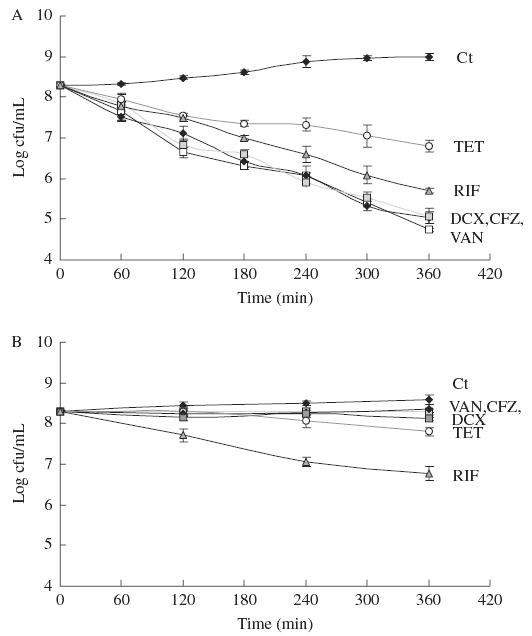
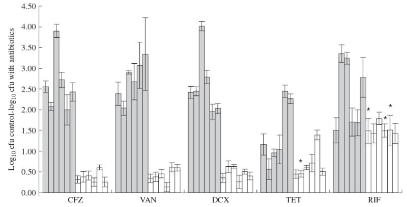
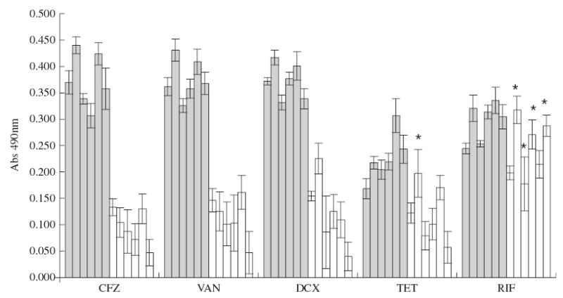
Methods: Several CoNS strains were grown planktonically or as biofilms to determine the effect of the mode of growth on the level of susceptibility to antibiotics with different mechanisms of action. The utility of a new, rapid colorimetric method that is based on the reduction of a tetrazolium salt (XTT) to measure cell viability was tested by comparison with standard bacterial enumeration techniques. A 6 h kinetic study was performed using dicloxacillin, cefazolin, vancomycin, tetracycline and rifampicin at the peak serum concentration of each antibiotic.
Results: In planktonic cells, inhibitors of cell wall synthesis were highly effective over a 3 h period. Biofilms were much less susceptible than planktonic cultures to all antibiotics tested, particularly inhibitors of cell wall synthesis. The susceptibility to inhibitors of protein and RNA synthesis was affected by the biofilm phenotype to a lesser degree. Standard bacterial enumeration techniques and the XTT method produced equivalent results both in biofilms and planktonic assays.
Conclusions: This study provides a more accurate comparison between the antibiotic susceptibilities of planktonic versus biofilm populations, because the cell densities in the two populations were similar and because we measured the concentration required to inhibit bacterial metabolism rather than to eradicate the entire bacterial population. While the biofilm phenotype is highly resistant to antibiotics that target cell wall synthesis, it is fairly susceptible to antibiotics that target RNA and protein synthesis.
Figures
References
-
- Voung C, Otto M. Staphylococcus epidermidis infections. Microbes Infect. 2002;4:481–9. - PubMed
-
- Costerton J, Stewart P, Greenberg P. Bacterial Biofilms: a commom cause of persistent infections. Science. 1999;284:1318–22. - PubMed
-
- Arciola C, Campoccia D, Montanaro L. Effects on antibiotic resistance of Staphylococcus epidermidis following adhesion to polymethyl-methacrylate and to silicone surfaces. Biomaterials. 2002;23:1495–1502. - PubMed
-
- Monzón M, Oteiza C, Leiva J, et al. Biofilm testing of Staphylococcus epidermidis clinical isolates: low performance of vancomycin in relation to other antibiotics. Diagn Microbiol Infect Dis. 2002;44:319–24. - PubMed
-
- Jansen B, Kristinsson K, Jansen S, et al. In-vitro efficacy of a central venous catheter complexed with iodine to prevent bacterial colonization. J Antimicrob Chemother. 1992;30:135–9. - PubMed
Publication types
MeSH terms
Substances
Grants and funding
LinkOut - more resources
Full Text Sources
Other Literature Sources
Medical
