. 2005 Sep;89(3):1893-901.
doi: 10.1529/biophysj.105.064212. Epub 2005 Jun 24.

Caveolin-3 is adjacent to a group of extradyadic ryanodine receptors

Affiliations

Caveolin-3 is adjacent to a group of extradyadic ryanodine receptors

David R L Scriven et al. Biophys J. 2005 Sep.

Abstract

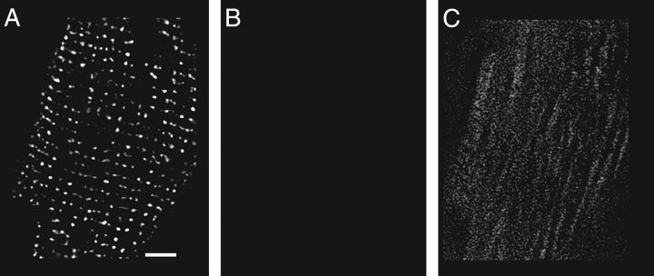
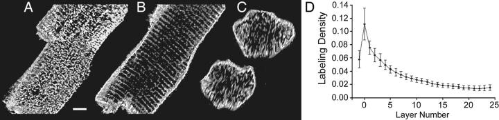
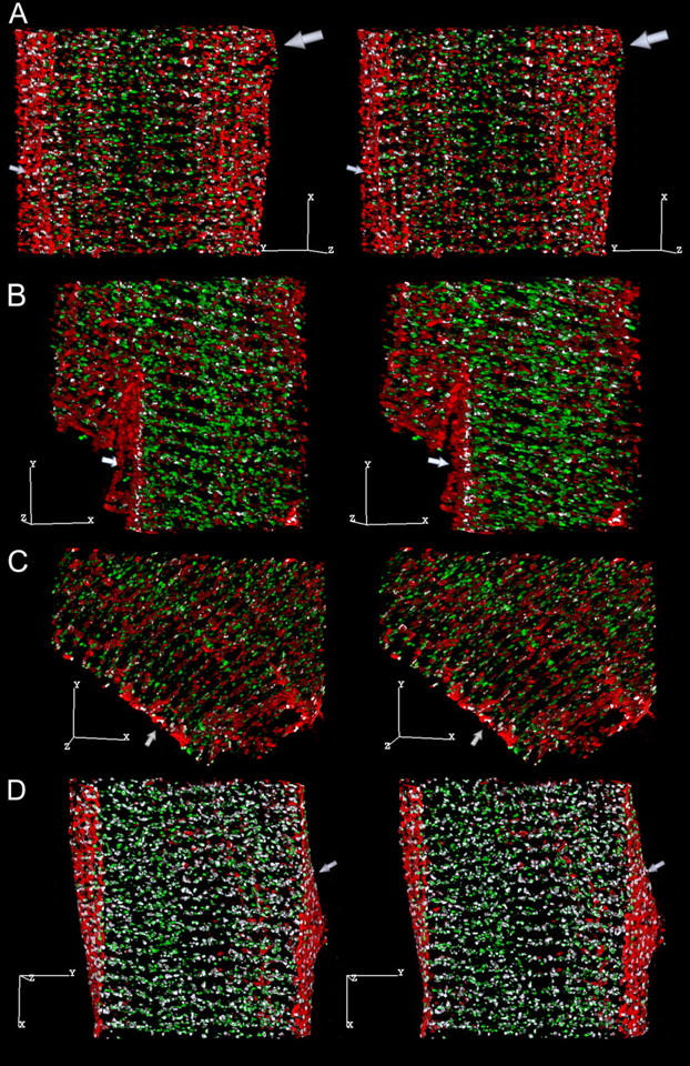
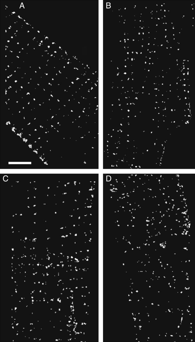
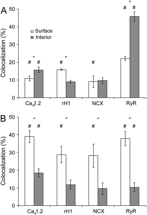
Caveolae are present in almost all cells and concentrate a wide variety of signaling molecules, receptors, transporters, and ion pumps. We have investigated the distribution of the ryanodine receptor, the Na(+)/Ca(2+) exchanger, the predominant Na(+) channel isoform rH1, and the L-type calcium channel, Ca(v)1.2, relative to the muscle-specific caveolin isoform, caveolin-3, in adult rat ventricular myocytes. Three-dimensional immunofluorescence images were deconvolved and analyzed. Caveolin-3 colocalizes with all of these molecules at the surface of the cell, but there is no significant colocalization between caveolin-3 and either the Na(+)/Ca(2+) exchanger or the Na(+) channel in the cell interior. The distribution of the surface colocalization indicates that the caveolae that colocalize with each molecule form distinct populations. This organization indicates that there are multiple populations of caveolae separable by location and occupants. In the interior of the cell, caveolin-3 shows a marked colocalization with a population of ryanodine receptors that are separate from those within the dyad. Because of their location, the signaling molecules contained within these caveolae may have preferred access to the neighboring nondyadic ryanodine receptors.


Figures

FIGURE 1
FIGURE 1
Labeling with two monoclonal antibodies. Each panel is a single image plane showing the distribution of RyR. (A) Distribution detected by a FITC-tagged anti-rabbit IgG antibody, after the monoclonal anti-RyR antibody had been transformed with a mouse anti-rabbit Fab fragment. Scale bar is 5 μm. (B) The same cell labeled with a Texas Red-tagged anti-mouse IgG and displayed identically to A shows nothing. (C) Image B, displayed 10× brighter, shows only nonspecific labeling.
FIGURE 2
FIGURE 2
Fitting the cell surface and generating the internal layers. (A) RyR distribution (image is 23 × 34 × 23 μm). Scale bar is 5 μm in each dimension. (B) Image in A, rotated about the x and y axes as indicated. (C) Image B, with the fitted surface. (D) The fitted surface from C and an interior layer (No. 15). (E) Two 1-μm-thick segments isolated from D.
FIGURE 3
FIGURE 3
Distribution of caveolin-3. The image is 47 × 50 × 16 μm, and the scale bar is 5 μm. (A) Caveolin-3 on the myocyte surface. Image is 4 μm deep. (B) The middle section (3 μm deep) shows that caveolin-3 in the interior is on or near the Z-lines and that the labeling intensity is dimmer than the surface. (C) Two cross sections (1 μm deep). D. Density of caveolin-3 labeling as a function of distance from the cell surface (N = 28).
FIGURE 4
FIGURE 4
Stereo pairs (6° rotation between images) showing the distribution of caveolin-3 (red) relative to: (A) Cav1.2 (green). The coincident voxels are white, and the scale bar is 5 μm in each dimension in this and all subsequent images. Image dimensions are 22 × 27 × 9.5 μm. There are 14,173 colocalized voxels: 27.5% of the Cav1.2 and 15.7% of the caveolin-3 voxels. The small arrow points to the cell surface, and the large arrow to a region near the surface where the majority of the colocalized voxels appear to be located. (B) NCX (green). Image dimensions are 21 × 22 × 9 μm. There are 8308 coincident voxels: 17.4% of the NCX and 7.8% of the caveolin-3 voxels. The arrow points to colocalized voxels located between the Z-lines on the cell surface. (C) The rH1 isoform of the Na+ channel (green). Image dimensions are 26 × 22 × 6 μm. There are 7785 coincident voxels: 26.2% of the Na+ channel and 10.6% of the caveolin-3 voxels. The arrow points to colocalized voxels between the Z-lines on the cell surface. (D) RyR (green). Image dimensions are 25 × 26 × 10.75 μm. There are 37,414 colocalized voxels: 37.5% of the RyR and 34.5% of the caveolin-3 voxels. The arrow points to a string of colocalized voxels at the Z-line on the cell surface.
FIGURE 5
FIGURE 5
Surface distribution showing colocalization of labeled proteins with caveolin-3. To produce the images, the coordinates of the surface were used to isolate a rind, 1 μm thick, from the cell surface. The resulting images were bisected in x, y to prevent overlap of the front and back surfaces, and only the colocalized voxels were displayed. The scale bar is 5 μm. (A) RyR. (B) Cav1.2. (C) NCX. (D) Na+ channel.
FIGURE 6
FIGURE 6
Histogram of colocalization at the surface and the interior of the cell. (A) The percentage of voxels labeled for caveolin-3 that also contained the indicated molecule B. The reverse: the percentage of voxels labeled for the indicated molecule that also contained caveolin-3. (★) denotes a significant difference between the surface and the interior (p < 0.05), (#) denotes a colocalization value significantly greater than predicted by chance (p < 0.05). The number of image pairs examined was Cav1.2 (N = 9); Na+ channel (N = 4); NCX (N = 4); and RyR (N = 11).
FIGURE 7
FIGURE 7
(A) Percentage of voxels labeled for RyR (▪) or Cav1.2 (□) that also contained caveolin-3. (B) The percentage of voxels labeled for caveolin-3 that also contained RyR (▪) or Cav1.2 (□). (C) Labeling density, i.e., number of lit voxels/total voxels, for RyR (▪) and Cav1.2 (□).

References

    1. Razani, B., S. E. Woodman, and M. P. Lisanti. 2002. Caveolae: from cell biology to animal physiology. Pharmacol. Rev. 54:431–467. - PubMed
    1. Parton, R. G. 1996. Caveolae and caveolins. Curr. Opin. Cell Biol. 8:542–548. - PubMed
    1. Song, K. S., P. E. Scherer, Z. Tang, T. Okamoto, S. Li, M. Chafel, C. Chu, S. Kohtz, and M. P. Lisanti. 1996. Expression of caveolin-3 in skeletal, cardiac, and smooth muscle cells. J. Biol. Chem. 271:15160–15165. - PubMed
    1. Rybin, V. O., P. W. Grabham, H. Elouardighi, and S. F. Steinberg. 2003. Caveolae-associated proteins in cardiomyocytes: caveolin-2 expression and interactions with caveolin-3. Am. J. Physiol. Heart Circ. Physiol. 285:H325–H332. - PubMed
    1. Hagiwara, Y., Y. Nishina, H. Yorifuji, and T. Kikuchi. 2002. Immunolocalization of caveolin-1 and caveolin-3 in monkey skeletal, cardiac and uterine smooth muscles. Cell Struct. Funct. 27:375–382. - PubMed

Publication types

MeSH terms

LinkOut - more resources