. 2005 Jul;138(3):1774-84.
doi: 10.1104/pp.105.061200. Epub 2005 Jun 24.

Dissecting defense-related and developmental transcriptional responses of maize during Ustilago maydis infection and subsequent tumor formation

Affiliations

Dissecting defense-related and developmental transcriptional responses of maize during Ustilago maydis infection and subsequent tumor formation

Christoph W Basse. Plant Physiol. 2005 Jul.

Abstract

Infection of maize (Zea mays) plants with the smut fungus Ustilago maydis triggers the formation of tumors on aerial parts in which the fungal life cycle is completed. A differential display screen was performed to gain insight into transcriptional changes of the host response. Some of the genes strongly up-regulated in tumors showed a pronounced developmental expression pattern with decreasing transcript levels from basal to apical shoot segments, suggesting that U. maydis has the capacity to extend the undifferentiated state of maize plants. Differentially expressed genes implicated in secondary metabolism were Bx1, involved in biosynthesis of the cyclic hydroxamic acid 2,4-dihydroxy-7-methoxy-2H-1,4-benzoxazin-3-one, and a novel putative sesquiterpene cyclase gene U. maydis induced (Umi)2. Together with the up-regulation of Umi11 encoding a cyclotide-like protein this suggests a nonconventional induction of plant defenses. Explicitly, U. maydis was resistant to 2,4-dihydroxy-7-methoxy-2H-1,4-benzoxazin-3-one but susceptible to its benzoxazolinone derivative 6-methoxy-2-benzoxazolinone. Infection studies of isolated leaves with U. maydis and Colletotrichum graminicola provided evidence for coregulation of Umi2 and PR-1 gene expression, with mRNA levels strongly determined by the extent of fungal colonization within tissue. However, in contrast to Umi2, transcript levels of PR-1 remained low in plants infected with wild-type U. maydis but were 8-fold elevated upon infection with an U. maydis mutant strongly attenuated in pathogenic development. This suggests that U. maydis colonization in planta suppresses a classical defense response. Furthermore, comparative expression analysis uncovered distinct transcriptional programs operating in the host in response to fungal infection and subsequent tumor formation.


Figures
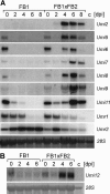
Figure 1.
Figure 1.
Kinetics of Umi/Umr gene expression. A, RNA-blot analysis of various maize genes identified by differential display analysis. Maize seedlings were inoculated with either FB1 or a mixture of FB1/FB2 sporidia. RNA was isolated from tissue immediately after inoculation (0) or after 2, 4, 6, and 8 d (numbers on top). Tissue from mock-infected plants and infected plants prior to tumor formation was harvested from leaf blades below the inoculation site. Lane c, Control RNA from dikaryotic hyphae (strain mixture FB1/FB2). Each gel was loaded with the same RNA preparations. B, RNA-blot analysis for Umi12. Identical amounts of the same samples (samples 8 dpi were omitted) as performed in A were loaded. A and B, Staining with methylene blue reflects the amounts of total RNA loaded (see Table I for quantification of signals).
Figure 2.
Figure 2.
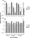
Developmental influence on Umi gene expression. A, Expression analysis of Umi6, 8, and 11. B, Expression analysis of Umi2, 7, and MAc1. A and B, Samples were taken from 5-d-old (1–3), 9-d-old (4–6), and 12-d-old seedlings (7–11) for isolation of total RNA. The basal-most 2 cm of the shoot from which the coleoptile leaf had been removed (1, 4, 7), 2 to 4 cm from the shoot base (2, 5, 8), 4 to 7 cm from the shoot base (9), leaf blade tissue of the first and second leaves (3), leaf blade tissue of the second and third leaves (6, 10), leaf blade tumor tissue of the second and third leaves 6 dpi (strains FB1/FB2; 11). Bars reflect relative expression levels individually calculated for each gene from the signals obtained by RNA-blot analysis. The sum of all bars assigned to a single gene corresponds to 100% (log scale). Umi11 and 2 (white bars, left), Umi8 and 7 (gray bars, middle), Umi6 and MAc1 (black bars, right).
Figure 3.
Figure 3.
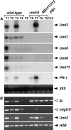
Expression of Umi genes related to fungal colonization and tumor induction. Maize seedlings were inoculated either with mixtures of strains FB1/FB2 (wild type, T1–T4), FB1Δmrb1/FB2Δmrb1 (Δmrb1, T6, and T7), AB311/AB312 (T8 and T9), or strain FB1 (T10). All samples were isolated from leaf blade tissue of the third and fourth leaves either 6 dpi or from untreated control plants of the same age (T5). T1, Leaf tumor tissue; T2, green leaf tissue lateral to tumors showing no signs of infection; T3, T7, and T9, tissue 2 to 5 cm below the inoculation site; T4, green leaf tissue distal (>2 cm) from tumors; T6, T8, and T10, tissue around (<1 cm) the inoculation site. A, RNA-blot analysis of Umi2, 6, 7, 8, 11, and PR-1. Staining with methylene blue reflects the amounts of total RNA loaded. B, RT-PCR analysis of U. maydis genes ip, mig2-5 (white triangles) and of maize genes Umi2 and H2B (control for integrity of RNA in all reactions). Number of cycles performed: 28 for mig2-5, Umi2, and H2B; 32 for ip.
Figure 4.
Figure 4.
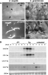
Defense-related gene expression in response to U. maydis and C. graminicola infection. A, Hyphal growth within plant tissue and C. graminicola penetration structures. Pictures were taken from U. maydis infected tissue 5 d after application of sporidia (JF1) and from C. graminicola infected tissue 1 d (bottom) and 3 d (top) after application of conidia (Cg). Maize tissue samples were assayed by epifluorescence (GFP; exposure time = 2 s) and/or differential interference contrast light microscopy (DIC). C. graminicola infection structures were stained with CBE. Hyphae and appressoria are marked by arrowheads. Bars = 10 μm. B, Time course of Umi and PR-1 expression during fungal infection. For each time point, RNA was isolated from three leaves either untreated or identically treated with U. maydis strain JF1 (JF1) and C. graminicola (Cg), respectively. Numbers on top refer to days after fungal application. Staining with methylene blue reflects the amounts of total RNA loaded.
Figure 5.
Figure 5.
Effect of cyclic hydroxamic acids on colony growth of U. maydis. Growth of U. maydis strains FB1 and FB2 on solid PD medium amended with various concentrations of DIMBOA (D), MBOA (M), and Trp (T) after 24 h. The final concentration of these compounds is indicated on the left. Trp was included as control indole compound in this assay.
Figure 6.
Figure 6.
Disulfide bridge arrangement of cyclotide domains and sequence comparison with UMI11. The putative cyclotide domain deduced from the Umi11 ORF (amino acid positions 56–88) was aligned with those of the mature Kalata B5 (subfamily 1), cycloviolacin O10 (subfamily 1), and cycloviolacin O12 (subfamily 2) proteins. The six highly conserved Cys residues are shaded black. Lines (top) refer to internal disulfide bridges. The extended line (bottom) refers to the ring closure between Ile and Pro. Identical or highly similar (Ile/Leu, Ser/Thr, Arg/Lys) positions dominating a column are shaded gray. Gaps were introduced to maximize the alignment.
Figure 7.
Figure 7.
Proposed scheme for distinct signaling pathways during U. maydis infection. Expression of Umi2 is strongly induced during hyphal colonization within plant tissue (arrows) and weakly by intimate contacts on the surface (dashed arrow). Umi2 and PR-1 are at least in part coregulated and not specific for U. maydis infection. Expression of Umi7 is influenced by hyphal growth within plant tissue and developmental signals as triggered during U. maydis colonization. Induced expression of Umi6, 8, and 11 results from developmental responses to U. maydis infection. White arrow heads refer to defense-related functions, while remaining Umi genes might have functions in fungal support. See text for further details.

References

    1. Altschul SF, Madden TL, Schäffer AA, Zhang J, Zhang Z, Miller W, Lipman DJ (1997) Gapped BLAST and PSI-BLAST: a new generation of protein database search programs. Nucleic Acids Res 25: 3389–3402 - PMC - PubMed
    1. Banuett F, Herskowitz I (1989) Different a alleles of Ustilago maydis are necessary for maintenance of filamentous growth but not for meiosis. Proc Natl Acad Sci USA 86: 5878–5882 - PMC - PubMed
    1. Banuett F, Herskowitz I (1996) Discrete developmental stages during teliospore formation in the corn smut fungus, Ustilago maydis. Development 122: 2965–2976 - PubMed
    1. Basse CW, Kerschbamer C, Brustmann M, Altmann T, Kahmann R (2002. b) Evidence for a Ustilago maydis steroid 5alpha-reductase by functional expression in Arabidopsis det2-1 mutants. Plant Physiol 129: 717–732 - PMC - PubMed
    1. Basse CW, Kolb S, Kahmann R (2002. a) A maize-specifically expressed gene cluster in Ustilago maydis. Mol Microbiol 43: 75–93 - PubMed

Publication types

MeSH terms