. 2005 Jul;46(7):2487-94.
doi: 10.1167/iovs.04-1333.

Leukocyte diapedesis in vivo induces transient loss of tight junction protein at the blood-retina barrier

Affiliations

Leukocyte diapedesis in vivo induces transient loss of tight junction protein at the blood-retina barrier

Heping Xu et al. Invest Ophthalmol Vis Sci. 2005 Jul.

Abstract

Purpose: Although much is now understood about the molecular structure of tight junctions (TJs), little is known about the regulation of their function during neural inflammatory disease processes in vivo. The mechanisms by which leukocytes transmigrate the blood-retina barrier (BRB) without affecting endothelial permeability are controversial.

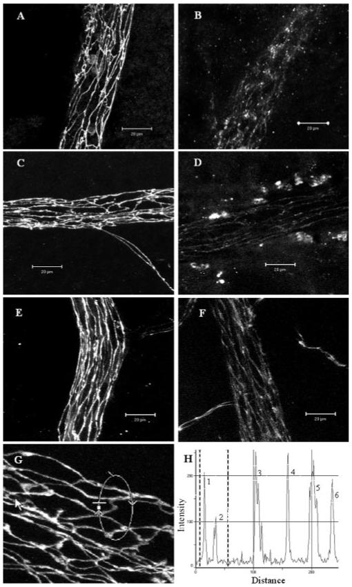
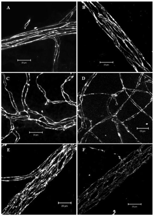
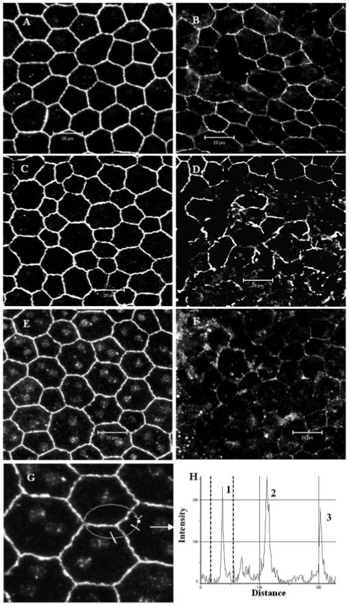
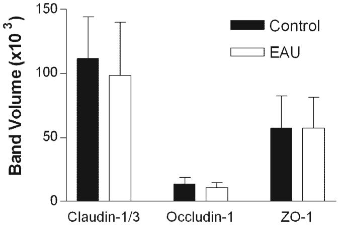
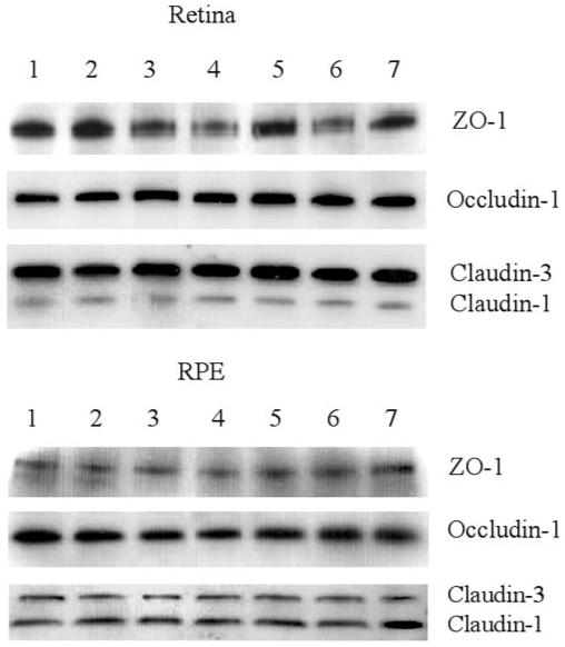
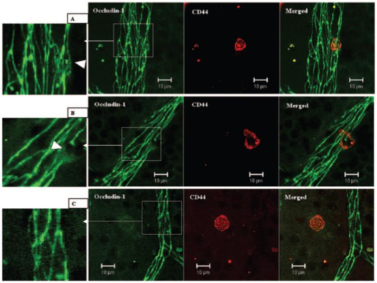
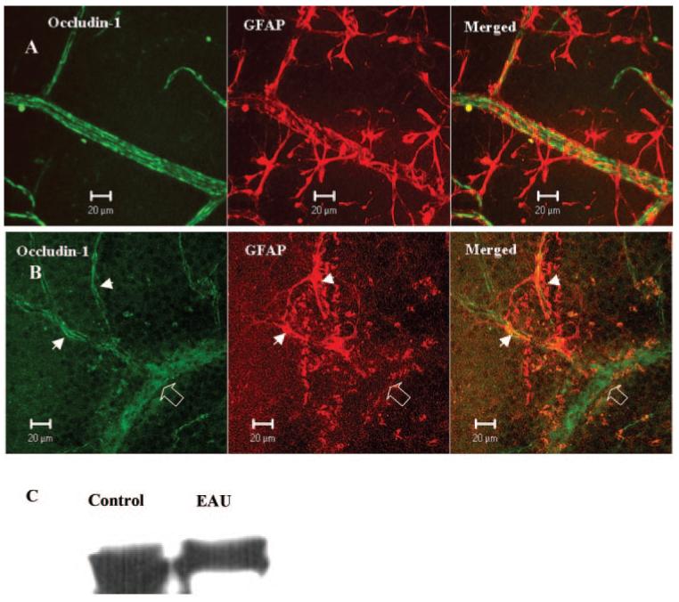
Methods: Confocal immunofluorescence microscopy of ex vivo retinal wholemounts was used to study BRB integrity during leukocyte adhesion and migration during experimental autoimmune uveoretinitis (EAU). Western blot analysis was used to measure levels of TJ proteins in EAU retina and RPE and in normal retina or RPE cultured with cytokines or chemokines.

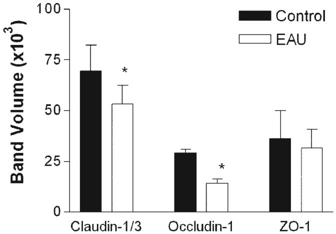
Results: No evidence for discontinuity or other weakness of the endothelial or epithelial barrier at tricellular corners was observed, and maximum disruption of TJ protein expression was focused in retinal venules correlating with sites of leukocyte extravasation. Areas of maximum TJ protein loss in vivo also correlated with redistribution or loss of ensheathing astrocyte processes on venules but not adjacent capillaries or arterioles. Exposure of normal choroidal and retinal explants ex vivo to cytokines and chemokines alone did not downregulate total occludin-1 or claudin-3 TJ protein expression.

Conclusions: The data presented herein support an active role for leukocytes in TJ disruption and blood-retina barrier (BRB) breakdown during retinal inflammation and further implicate venule microenvironment as a key factor in leukocyte recruitment to retinal tissue in vivo.


Figures

Figure 1
Figure 1
Confocal images of TJ protein expression in retinal venules of normal and EAU mice. Retinal wholemounts from normal nonimmunized B10R.III mice (A, C, E, G) or day 12 pi mice (B, D, F) were stained with anti claudin-1/3 (A, B), anti occludin-1 (C, D, G), and anti ZO-1 (E, F). All TJ proteins were evident at the interfaces between adjacent endothelial cells of retinal vessels from normal B10R.III mice and only weakly appeared in inflamed venules. (H) Histogram of fluorescence intensity of occludin-1 along the circular track shown in (G). Peaks 1 and 2 are two junction sites within the marked area in (G). Peaks 3, 4, and 6 are three tricellular corner sites. Fluorescence intensity of occludin-1 was not reduced in the tricellular areas. Images shown are reconstructions of a series of Z-stacks (15-30-μm thickness) of retinal venule segments. Bars, 20 μm.
Figure 2
Figure 2
Confocal images of TJ protein occludin-1 in retinal vessels of normal and EAU mice. (A, B) arterioles; (C, D) capillaries; (E, F) venules. (A, C, E) Normal control; (B, D, F) EAU day 12 pi. Note the remarkable reduction of occludin-1 expression in retinal venules of EAU mouse. Images shown are reconstructions of a series of Z-stacks (15-30 μm thickness). Bars, 20 μm.
Figure 3
Figure 3
Western blot analysis for TJ proteins in retinas from normal and EAU mice. Retinas were dissected from the choroids-RPE tissue in normal or EAU mice (C57BL/6 milder disease) and proteins extracted for Western blot analysis with claudin-1/3, occludin-1, and ZO-1. Data shown are expressed as the mean ± SEM. n = 3 mice, *P < 0.05 Student’s t-test.
Figure 4
Figure 4
Confocal images of TJ protein expression in RPE cells of normal and EAU mice. Wholemounts of choroids (include RPE) from normal nonimmunized B10R.III mice (A, C, E, G) or day 12 pi mice (B, D, F) were stained with anti claudin-1/3 (A, B), anti occludin-1 (C, D), and anti ZO-1 (E, F, G). All TJ proteins were detected around the entire circumference of RPE cells from normal B10R.III mice and markedly disrupted in inflamed RPE cells. (H) Histogram of fluorescence intensity of ZO-1 along the circular track shown in (G). Peaks 1 and 3 are two cell border sites and peak 2 is the tricellular corner site. Images are reconstructions of a series of Z-stacks (12-μm thickness). Bars, 20 μm.
Figure 5
Figure 5
Quantification of results in a Western blot analysis of TJ proteins in RPE cells from normal and EAU mice. RPE-choroid tissues were dissected from normal or EAU mice (C57BL/6 milder disease) and proteins extracted for Western blot analysis with claudin-1/3, occludin-1, and ZO-1. Data are expressed as the mean ± SEM; n = 3 mice.
Figure 6
Figure 6
Western blot analysis of ex vivo retinas and RPE cells for TJ proteins ZO-1, occludin-1, and claudin-1/3 after cytokine or chemokine treatment. Retinal and choroid-RPE tissues were dissected from normal mouse eyes and incubated for 24 hours in the presence or absence of different cytokines or chemokines. Proteins were extracted for Western blot analysis. Row 1, control; row 2, IFN-γ; row 3, TNF-α; row 4, IL-1β; row 5, MCP-1; row 6, MIP-1α; and row 7, RANTES. Data are representative of three experiments. Bands representing either claudin-1 (20 kDa) or claudin-3 (22 kDa) were differentiated in blots, by using specific claudin-1 and claudin-3 antibodies that became available during the period of the study (data not shown).
Figure 7
Figure 7
Changes of occludin-1 in retinal venules during leukocyte adhesion and transendothelial cell migration. Day 9 pi EAU mice were gently perfused with PBS. Retinal wholemounts were double stained with anti-occludin-1 (FITC) and anti-CD44 (R-PE). (A) One CD44+ leukocyte adhering to a retinal venule. TJ protein occludin-1 is diffuse in the adhering area (inset, arrowhead). (B) One CD44+ leukocyte was in the process of transendothelial cell migration. TJ protein occludin-1 was absent in the transmigration area and diffuse in the adjacent area (inset, arrowhead). (C) One CD44+ cells had migrated into the tissue, and there was no occludin-1 disruption in surrounding vessels. Images are reconstructions from a series of Z-stacks (15-40-μm thickness). Bars, 10 μm.
Figure 8
Figure 8
Occludin-1 and GFAP immunoreactivity in normal and EAU mouse retina. Retinal wholemounts from a normal B10R.III mouse (A) and a day 12 pi EAU mouse (B) were stained with anti-occludin-1 and anti-GFAP. Samples were observed by confocal microscopy. Note the total loss of GFAP staining in the vessel segment with occludin-1 disruption (diffuse occludin-1 staining, open arrow) and the preservation of occludin-1 in capillaries where GFAP astrocytes are maintained intact (arrowheads). Images shown are reconstructions from a series of Z-stacks (40-μm thickness). (C) Western blot of retinal tissue from control and EAU mice showing reduced GFAP protein expression in EAU retina.

References

    1. Xu H, Manivannan A, Liversidge J, Sharp PF, Forrester JV, Crane IJ. Requirements for passage of T lymphocytes across non-inflamed retinal microvessels. J Neuroimmunol. 2003;142:47–57. - PubMed
    1. Xu H, Forrester JV, Liversidge J, Crane IJ. Leukocyte trafficking in experimental autoimmune uveitis: breakdown of blood-retinal barrier and upregulation of cellular adhesion molecules. Invest Ophthalmol Vis Sci. 2003;44:226–234. - PubMed
    1. Wolburg H, Lippoldt A. Tight junctions of the blood-brain barrier: development, composition and regulation. Vasc Pharmacol. 2002;38:323–337. - PubMed
    1. Barber AJ, Antonetti DA, Gardner TW, the Penn State Retina Research Group Altered expression of retinal occludin and glial fibrillary acidic protein in experimental diabetes. Invest Ophthalmol Vis Sci. 2000;41:3561–3568. - PubMed
    1. Morcos Y, Hosie MJ, Bauer HC, Chan-Ling T. Immunolocalization of occludin and claudin-1 to tight junctions in intact CNS vessels of mammalian retina. J Neurocytol. 2001;30:107–123. - PubMed

Publication types

MeSH terms