

Antibacterial properties of some cyclic organobismuth(III) compounds
- PMID: 15980343
- PMCID: PMC1168658
- DOI: 10.1128/AAC.49.7.2729-2734.2005
Antibacterial properties of some cyclic organobismuth(III) compounds
Abstract
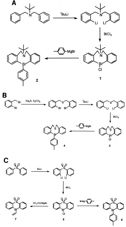
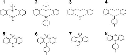
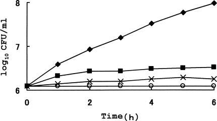
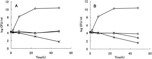
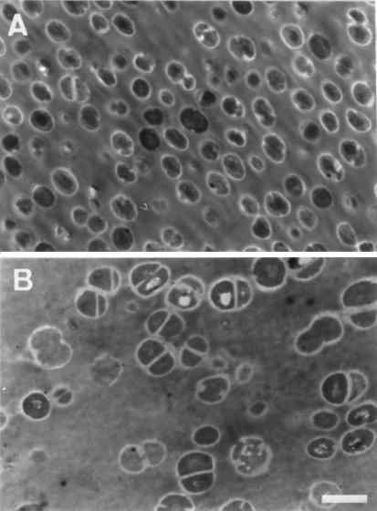
Bismuth compounds are known for their low levels of toxicity in mammals, and various types of bismuth salts have been used to treat medical disorders. As part of our program to probe this aspect of bismuth chemistry, cyclic organobismuth compounds 1 to 8 bearing a nitrogen or sulfur atom as an additional ring member have been synthesized, and their antimicrobial activities against five standard strains of gram-negative and gram-positive bacteria were assessed. The eight-membered-ring compounds, compounds 1 to 3, exhibited MICs of less than 0.5 microg/ml against Staphylococcus aureus and were more active than the six-membered ones, compounds 5 to 8 (MICs, 4.0 to 16 microg/ml). The gram-positive bacteria (Staphylococcus aureus, Bacillus subtilis, and Enterococcus faecalis) were more susceptible to both types of ring compounds than the gram-negative ones (Escherichia coli and Pseudomonas aeruginosa). Treatment with polymyxin B nonapeptide increased the susceptibility of E. coli to cyclic organobismuth compounds, indicating the low permeability of the outer membrane of gram-negative bacteria to the compounds. Compound 1 also had activity against methicillin-resistant S. aureus, which had an MIC for 90% of the hospital stock strains of 1.25 microg/ml. The killing curves for S. aureus treated with compound 1 or 3 revealed a static effect at a low dose (2x the MIC). However, when S. aureus was treated with 10x the MIC of compound 1 or 3, there was an approximately 3-log reduction in the viable cell number after 48 h of treatment. Electron microscopic inspection demonstrated a considerable increase in the size of S. aureus and the proportion of cells undergoing cell division after treatment with compound 1 at 0.5x the MIC.
Figures
References
-
- Bierer, D. W. 1990. Bismuth subsalicylate: history, chemistry, and safety. Rev. Infect. Dis. 12(Suppl. 1):S3-S8. - PubMed
-
- Briand, G. G., and N. Burford. 1999. Bismuth compounds and preparations with biological or medicinal relevance. Chem. Rev. 99:2601-2657. - PubMed
-
- Cornick, N. A., M. Silva, and S. L. Gorbach. 1990. In vitro antibacterial activity of bismuth subsalicylate. Rev. Infect. Dis. 12(Suppl. 1):S9-S10. - PubMed
-
- Dittes, U., E. Vogel, and B. K. Keppler. 1997. Overview on bismuth(III) and bismuth(V) complexes with activity against Helicobacter pylori. Coord. Chem. Rev. 163:345-364.
MeSH terms
Substances
LinkOut - more resources
Full Text Sources
Other Literature Sources
Medical
Molecular Biology Databases
