. 2005 Sep;146(1):41-8.
doi: 10.1038/sj.bjp.0706312.

The hypoglycaemic activity of fenugreek seed extract is mediated through the stimulation of an insulin signalling pathway

Affiliations

The hypoglycaemic activity of fenugreek seed extract is mediated through the stimulation of an insulin signalling pathway

Maleppillil Vavachan Vijayakumar et al. Br J Pharmacol. 2005 Sep.

Abstract

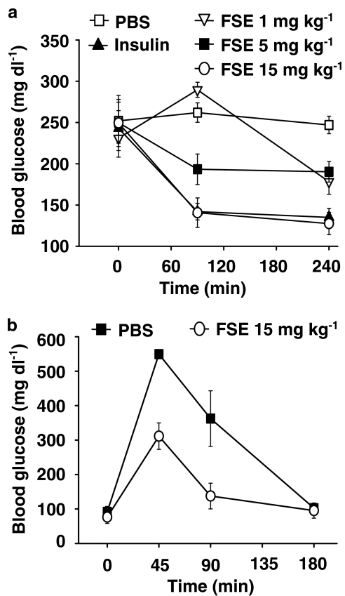
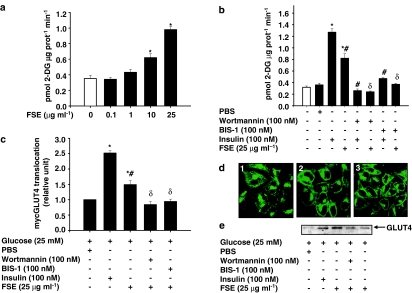
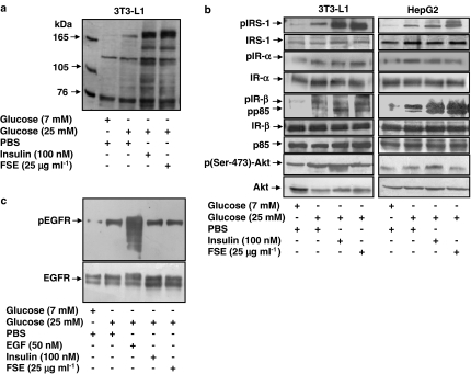
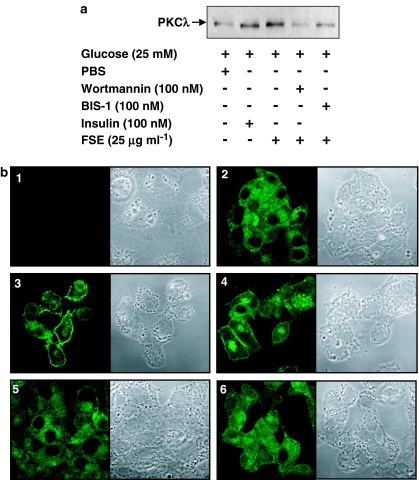
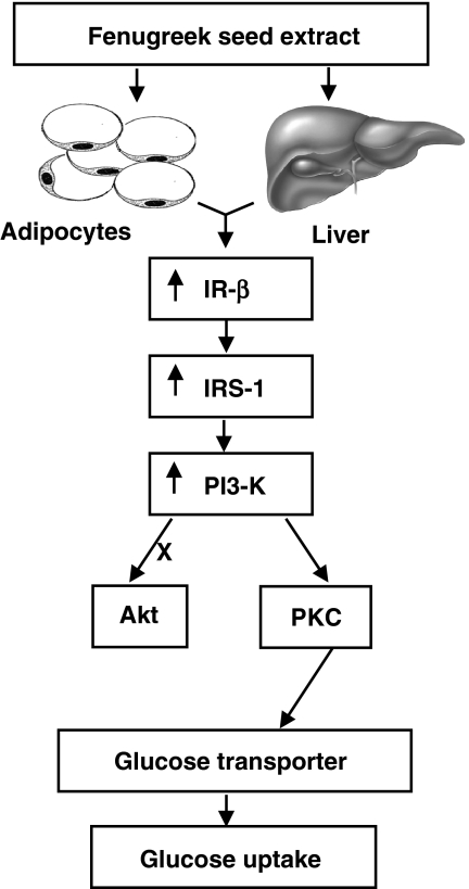
The in vivo hypoglycaemic activity of a dialysed fenugreek seed extract (FSE) was studied in alloxan (AXN)-induced diabetic mice and found to be comparable to that of insulin (1.5 U kg(-1)). FSE also improved intraperitoneal glucose tolerance in normal mice. The mechanism by which FSE attenuated hyperglycaemia was investigated in vitro. FSE stimulated glucose uptake in CHO-HIRc-mycGLUT4eGFP cells in a dose-dependent manner. This effect was shown to be mediated by the translocation of glucose transporter 4 (GLUT4) from the intracellular space to the plasma membrane. These effects of FSE on GLUT4 translocation and glucose uptake were inhibited by wortmannin, a phosphatidylinositol 3-kinase (PI3-K) inhibitor, and bisindolylmaleimide 1, a protein kinase C (PKC)-specific inhibitor. In vitro phosphorylation analysis revealed that, like insulin, FSE also induces tyrosine phosphorylation of a number of proteins including the insulin receptor, insulin receptor substrate 1 and p85 subunit of PI3-K, in both 3T3-L1 adipocytes and human hepatoma cells, HepG2. However, unlike insulin, FSE had no effect on protein kinase B (Akt) activation. These results suggest that in vivo the hypoglycaemic effect of FSE is mediated, at least in part, by the activation of an insulin signalling pathway in adipocytes and liver cells.


Figures

Figure 1
Figure 1
In vivo hypoglycaemic activity of FSE. (a) AXN-induced diabetic Swiss albino mice were injected (i.p.) with vehicle (PBS), insulin or FSE (1, 5 or 15 mg kg−1). FSE at a dose of 15 mg kg−1 produced a significant decrease in blood glucose levels compared with PBS control (P<0.01). (b) IPGTT in glucose-loaded (3 g kg−1) normal Swiss albino mice. FSE-injected mice had significantly lower levels of blood glucose (P<0.01 vs PBS treated) at 45 and 90 min after its administration.
Figure 2
Figure 2
Effects of FSE on glucose transport and GLUT4 translocation. (a) CHO-HIRc-mycGLUT4eGFP cells were incubated with the indicated concentrations of FSE for 30 min before 2-DG uptake was measured. (b) CHO-HIRc-mycGLUT4eGFP cells were preincubated in the absence or presence of either wortmannin or BIS-1 for 20 and 60 min, respectively, followed by treatment with insulin or FSE for 10 and 30 min, respectively, before 2-DG uptake was measured. Each value shown in (a) and (b) represents the mean and s.e.m. of three independent experiments performed in triplicate (*P<0.05 vs basal; #P<0.05 vs insulin control; δP<0.05 vs FSE control). The open column represents the basal values. (c) Differentiated 3T3-L1-mycGLUT4 cells were serum-starved in DMEM and incubated in the absence or presence of wortmannin or BIS-1 for 20 and 60 min, respectively, followed by treatment with insulin or FSE for 10 and 30 min, respectively, before mycGLUT4 translocation was measured. Data represent the mean and s.e.m. of triplicate experiments and are expressed as relative translocation compared with control, which was set at 1.0 (*P<0.05 vs basal value; #P<0.05 vs insulin control; δP<0.05 vs FSE control). (d) CHO-HIRc-mycGLUT4eGFP cells were serum-starved in DMEM for 3 h followed by treatment with (1) PBS (2) insulin or (3) FSE. Cells were fixed and processed for confocal microscopy analysis. Representative pictures of at least three independent experiments are shown. (e) 3T3-L1 adipocytes were incubated in the absence or presence of wortmannin or BIS-1 for 20 and 60 min, respectively, followed by treatment with insulin or FSE for 10 and 30 min, respectively. Plasma membranes were prepared as described in the Methods section and then subjected to immunoblot analysis with antibodies against GLUT4.
Figure 3
Figure 3
Effects of FSE on insulin signalling pathways. Differentiated 3T3-L1 adipocytes or HepG2 cells were serum-starved and incubated with insulin or FSE for 10 and 30 min, respectively. The cells were washed, lysed, subjected to SDS–PAGE followed by immunoblot analysis with phospotyrosine (PY-20) antibodies or other specific antibodies and developed by enhanced chemiluminescence. (a) Tyrosine phosphorylation of cellular proteins of differentiated 3T3-L1 adipocytes. (b) Phosphorylation of IRS-1, IR-α, IR-β, p85 and Akt in 3T3-L1 adipocytes and HepG2 cells. (c) Serum-starved A431 cells were treated with EGF, insulin or FSE and washed, lysed and subjected to immunoblot analysis. The results shown are representative of three independent experiments.
Figure 4
Figure 4
Effects of FSE on PKC translocation. (a) Serum-starved 3T3-L1 adipocytes were preincubated in the absence or presence of wortmannin or BIS-1 for 20 and 60 min, respectively, followed by treatment with FSE for 10 and 30 min, respectively. Plasma membranes were prepared as described in the Methods section and subjected to protein immunoblot analysis with antibodies against PKCλ. (b) HepG2 cells were serum-starved and preincubated in the absence or presence of wortmannin or BIS-1 for 20 and 60 min, respectively, followed by treatment with insulin or FSE for 10 and 30 min, respectively. Immunostaining was performed as described in the Methods section. (1) Negative control, (2) basal, (3) insulin, (4) FSE, (5) and (6) pretreated with wortmannin and BIS-1, respectively, followed by treatment with FSE. The results shown are representative of at least two independent experiments.
Figure 5
Figure 5
A proposed model for cellular effects of fenugreek seed extract on glucose homeostasis.

References

    1. ABDEL-BARRY J.A., ABDEL-HASSAN I.A., AL-HAKIEM M.H. Hypoglycemic and antihyperglycemic effects of Trigonella foenum-graecum leaf in normal and alloxan induced diabetic rats. J. Ethnopharmacol. 1997;58:149–155. - PubMed
    1. BASCH E., ULBRICHT C., KUO G., SZAPARY P., SMITH M. Therapeutic applications of fenugreek. Altern. Med. Rev. 2003;8:20–27. - PubMed
    1. CHEATHAM B., VLAHOS C.J., CHEATHAM L., WANG L., BLENIS J., KAHN C.R. Phosphatidylinositol 3-kinase activation is required for insulin stimulation of pp70 S6 kinase, DNA synthesis, and glucose transporter translocation. Mol. Cell. Biol. 1994;14:4902–4911. - PMC - PubMed
    1. CHHIPA R.R., SINGH S., SURVE S.V., VIJAYAKUMAR M.V., BHAT M.K. Doxycycline potentiates antitumor effect of cyclophosphamide in mice. Toxicol. Appl. Pharmacol. 2005;202:268–277. - PubMed
    1. CLANCY B.M., CZECH M.P. Hexose transport stimulation and membrane redistribution of glucose transporter isoforms in response to cholera toxin, dibutyryl cyclic AMP, and insulin in 3T3-L1 adipocytes. J. Biol. Chem. 1990;265:12434–12443. - PubMed

Publication types