. 2005 Jul 4;170(1):81-90.
doi: 10.1083/jcb.200412015. Epub 2005 Jun 27.

TGF-{beta} maintains dormancy of prostatic stem cells in the proximal region of ducts

Affiliations

TGF-{beta} maintains dormancy of prostatic stem cells in the proximal region of ducts

Sarah N Salm et al. J Cell Biol. .

Abstract

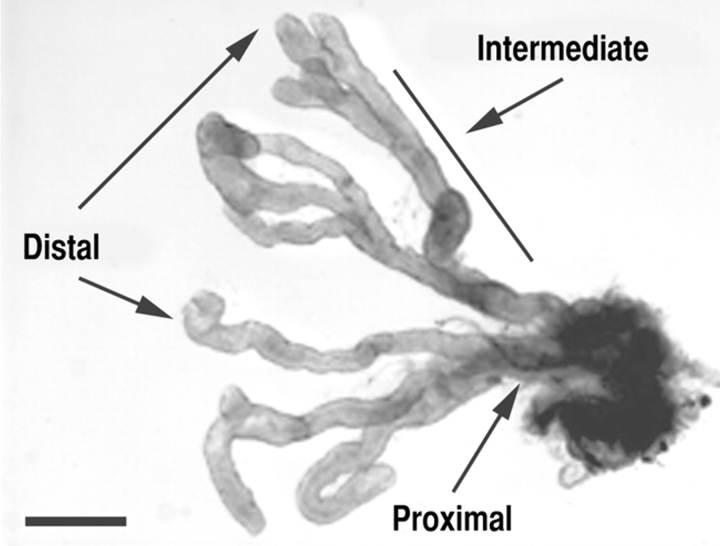
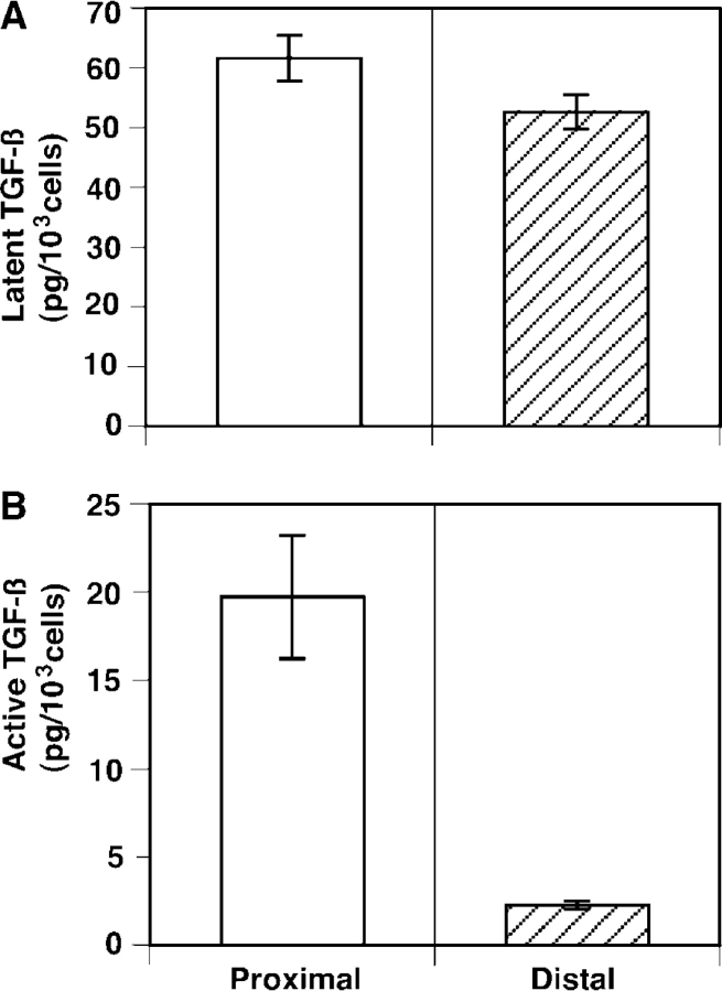
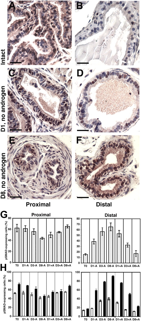
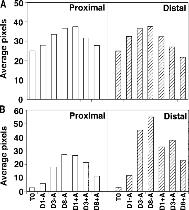
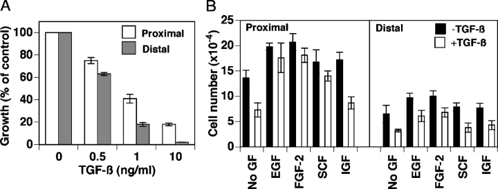
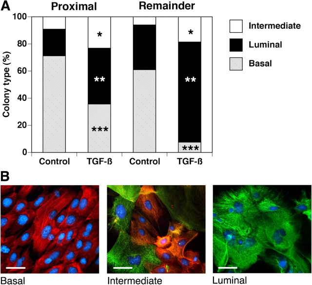
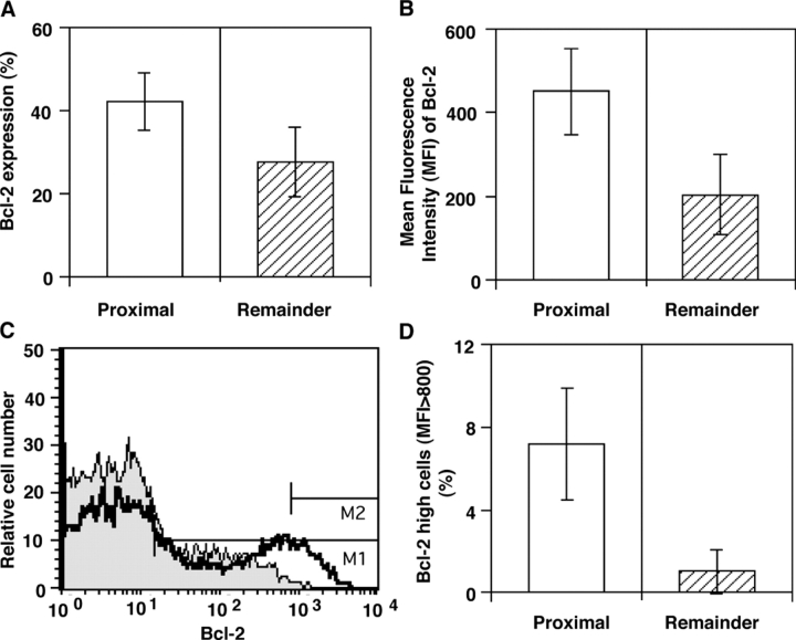
We have previously shown that prostatic stem cells are located in the proximal region of mouse prostatic ducts. Here, we show that this region responds differently to transforming growth factor (TGF)-beta than the distal ductal region and that under physiological conditions androgens and TGF-beta are crucial overall regulators of prostatic tissue homeostasis. This conclusion is supported by the observations showing that high levels of TGF-beta signaling are present in the quiescent proximal region of ducts in an androgen-replete animal and that cells in this region overexpress Bcl-2, which protects them from apoptosis. Moreover, androgen ablation reverses the proximal-distal TGF-beta signaling gradient, leading to an increase in TGF-beta signaling in the unprotected distal region (low Bcl-2 expression). This reversal of TGF-beta-mediated signaling accompanies apoptosis of cells in the distal region and gland involution after androgen withdrawal. A physiological TGF-beta signaling gradient (high proximally and low distally) and its functional correlates are restored after androgen replenishment. In addition to highlighting the regulatory role of androgens and TGF-beta, these findings may have important implications for the deregulation of the stem cell compartment in the etiology of proliferative prostatic diseases.


Figures

Figure 1.
Figure 1.
Microdissected prostatic ducts. A segment from a microdissected mouse dorsal prostate showing the distal, intermediate, and proximal regions of prostatic ducts. Bar, 1 mm.
Figure 2.
Figure 2.
Proximal cells produce and activate more TGF-β than distal cells. (A) Heat-activated conditioned medium from proximal and distal cells (4 × 10⁴ cells/24-well dish) was placed on TGF-β–responsive mink lung cells. The amount of total TGF-β was quantified by comparison with a standard curve. (B) Active TGF-β was measured directly by quantifying luciferase activity using a coculture assay in which either proximal or distal cells (4 × 10³ cells/well) were combined with TGF-β–responsive mink lung cells (5 × 10³ cells/well) in a 96-well plate. The presence of TGF-β in A and B was verified using TGF-β–neutralizing antibodies.
Figure 3.
Figure 3.
pSMAD 2 and 3 expression are differentially regulated by androgens in proximal and distal cells. (A–F) Paraffin sections of the proximal (A, C, and E) and distal (B, D, and F) regions of ducts examined immunohistochemically for pSMAD2/3-expressing cells in tissues removed from androgen-replete animals (A and B), and after androgen deprivation for 1 d (D1, no androgen; C and D) and 8 d (D8, no androgen; E and F). Bars, 100 μm. (G) Quantification of the number of nuclei positive for pSMAD2/3 expression in proximal and distal cells in the intact prostate (T0), the prostate 1 (D1-A), 3 (D3-A), and 8 d (D8-A) after castration and after 1 (D1+A), 3 (D3+A), and 8 d (D8+A) of androgen administration to animals with involuted prostates. (H) Quantification of the number of basal and luminal nuclei positive for pSMAD2/3 expression in proximal and distal cells in the intact prostate (T0), the prostate 1 (D1-A), 3 (D3-A), and 8 d (D8-A) after castration and after 1 (D1+A), 3 (D3+A), and 8 d (D8+A) of androgen administration to animals with involuted prostates. White bars, basal cells; black bars, luminal cells.
Figure 4.
Figure 4.
TβRI expression is regulated by androgens in proximal and distal cells. TβRI was detected using RT-PCR (A) and Western blot (B) in the intact prostate (T0), the prostate 1 (D1-A), 3 (D3-A), and 8 d (D8-A) after castration and after 1 (D1+A), 3 (D3+A), and 8 d (D8+A) of androgen administration to animals with involuted prostates.
Figure 5.
Figure 5.
The inhibitory effect of TGF-β on cell growth is opposed by EGF, FGF-2, and SCF. Cells from the proximal or distal region were seeded (5 × 10³ cells/well) on collagen and overlaid with medium containing TGF-β (0.5, 1, or 10 ng/ml; A) and either TGF-β alone (0.5 ng/ml) or TGF-β together with either EGF (10 ng/ml), FGF-2 (10 ng/ml), SCF (100 ng/ml), or IGF-1 (10 ng/ml) (B). Control wells received EGF, FGF-2, SCF, IGF-1, or none of these factors in the absence of TGF-β. Wells were cultured for 10 d, after which cells were enumerated. The y axis in A is normalized to cells that were not treated with TGF-β. The histograms of the proximal and distal cells in B cannot be directly compared with each other because of their differential responses to TGF-β. The comparisons should be made between the control (No GF) and experimental samples within each of the two histograms.
Figure 6.
Figure 6.
Proximal cells are more resistant to the differentiating-inducing effects of TGF-β than cells from the remaining regions of ducts. (A) Cells from the proximal and remaining ductal regions (2,000 cells/collagen-coated 8-well chamber slide) were cultured for 5–7 d and TGF-β (0.1 ng/ml) was added for 48 h after which colonies of >100 cells were counted and examined for evidence of basal and luminal cytokeratins. The data are plotted as the colony type (basal, luminal, or intermediate) as a percentage of the total number of colonies and are the average of four pooled experiments. *, 1.2-fold, P > 0.05 NS; **, 1.8-fold, P < 0.0001; ***, 4.7-fold, P < 0.001. (B) Colonies were examined immunohistochemically using antibodies to basal or luminal cytokeratins and appropriate Alexa Fluor 594 or 488 secondary fluorescent antibodies to determine those colonies that were comprised of basal (red), intermediate (both basal and luminal), or luminal (green) cells. Bars, 50 μm.
Figure 7.
Figure 7.
Bcl-2 is highly expressed by cells from the proximal region of prostatic ducts. (A) More cells in the proximal region of prostatic ducts express Bcl-2 than those from the remaining regions of ducts (P < 0.00001). (B) Cells in the proximal region express higher levels of Bcl-2 per cell, as is indicated by a higher mean fluorescence intensity (MFI) (P < 0.00001). (C) A representative histogram of Bcl-2 expression by cells from the proximal region (thick solid line; MFI = 599) and the remaining ductal regions (gray filled area; MFI = 157). The marker M1 is set such that <1% of control cells are included within this marker. The marker M2 delineates Bcl-2high cells with MFI > 800. (D) Cells expressing high levels of Bcl-2 (MFI > 800) are 7.1-fold enriched in the proximal region of ducts compared with those in the remaining ductal regions (P < 0.00001).
Figure 8.
Figure 8.
A model for the regional regulation of prostatic homeostasis. A schematic diagram showing that in an androgen-replete prostate high levels of active TGF-β and TGF-β–mediated signaling in the proximal stem cell niche maintain the quiescence of stem cells in this region. In the distal region, low levels of active TGF-β and TGF-β–mediated signaling permit the division of transit-amplifying (TA) cells in this region. After castration, the decrease in androgen levels with concurrent increased levels of TGF-β and TGF-β–mediated signaling distally, lead to apoptosis of cells in this region, with the resulting involution of the prostate gland. Simultaneously, the decrease in TGF-β–mediated signaling in the proximal region sensitizes the cells in this region to mitogenic signals. When androgens are readministered these cells respond to androgen-induced mitogenic cytokines (GFs), thus contributing to prostatic regeneration. Simultaneously, as the distal TGF-β signaling activity declines, the transit-amplifying cells in this region also divide, resulting in regeneration of the gland.

References

    1. Abe, M., J.G. Harpel, C.N. Metz, I. Nunes, D.J. Loskutoff, and D.B. Rifkin. 1994. An assay for transforming growth factor-beta using cells transfected with a plasminogen activator inhibitor-1 promoter-luciferase construct. Anal. Biochem. 216:276–284. - PubMed
    1. Adams, J.M., and S. Cory. 1998. The Bcl-2 protein family: arbiters of cell survival. Science. 281:1322–1326. - PubMed
    1. Beachy, P.A., S.S. Karhadkar, and D.M. Berman. 2004. Tissue repair and stem cell renewal in carcinogenesis. Nature. 432:324–331. - PubMed
    1. Bruckheimer, E.M., and N. Kyprianou. 2002. Bcl-2 antagonizes the combined apoptotic effect of transforming growth factor-beta and dihydrotestosterone in prostate cancer cells. Prostate. 53:133–142. - PubMed
    1. Cashman, J.D., A.C. Eaves, and C.J. Eaves. 1992. Granulocyte-macrophage colony-stimulating factor modulation of the inhibitory effect of transforming growth factor-beta on normal and leukemic human hematopoietic progenitor cells. Leukemia. 6:886–892. - PubMed

Publication types

MeSH terms

Substances