. 2005 Jul 4;202(1):33-45.
doi: 10.1084/jem.20050471. Epub 2005 Jun 27.

Promiscuous gene expression in thymic epithelial cells is regulated at multiple levels

Affiliations

Promiscuous gene expression in thymic epithelial cells is regulated at multiple levels

Jens Derbinski et al. J Exp Med. .

Abstract

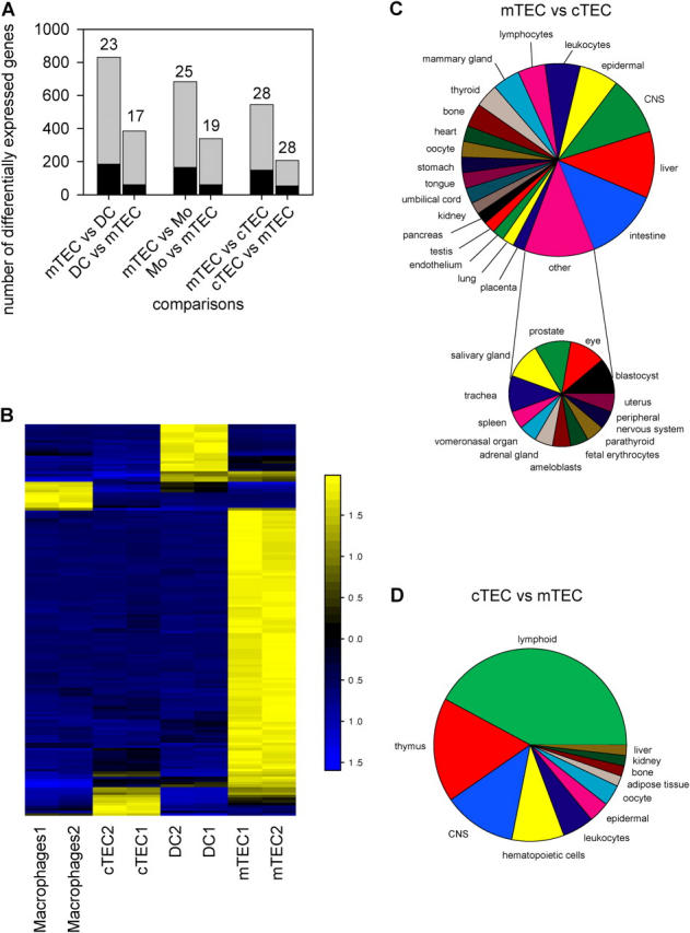
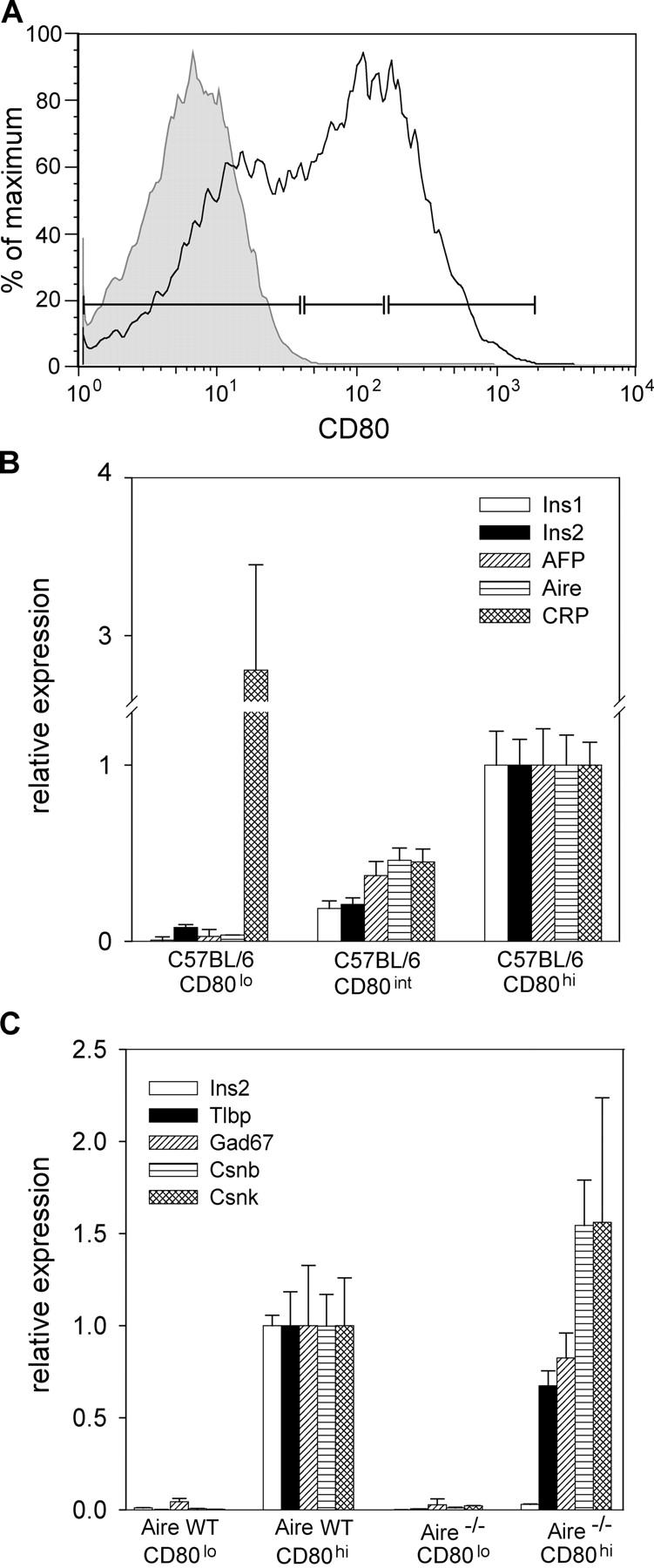
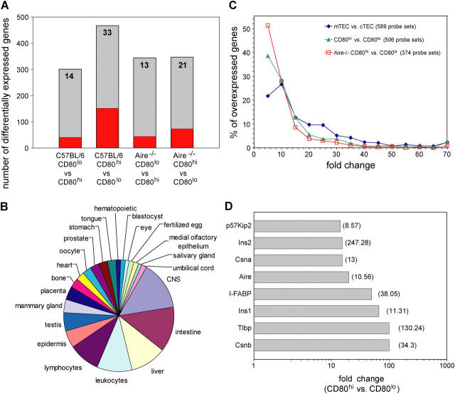
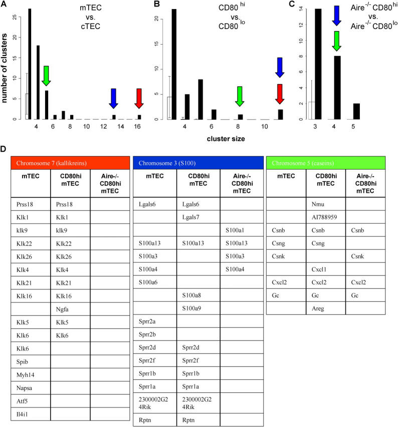
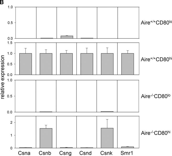
The role of central tolerance induction has recently been revised after the discovery of promiscuous expression of tissue-restricted self-antigens in the thymus. The extent of tissue representation afforded by this mechanism and its cellular and molecular regulation are barely defined. Here we show that medullary thymic epithelial cells (mTECs) are specialized to express a highly diverse set of genes representing essentially all tissues of the body. Most, but not all, of these genes are induced in functionally mature CD80(hi) mTECs. Although the autoimmune regulator (Aire) is responsible for inducing a large portion of this gene pool, numerous tissue-restricted genes are also up-regulated in mature mTECs in the absence of Aire. Promiscuously expressed genes tend to colocalize in clusters in the genome. Analysis of a particular gene locus revealed expression of clustered genes to be contiguous within such a cluster and to encompass both Aire-dependent and -independent genes. A role for epigenetic regulation is furthermore implied by the selective loss of imprinting of the insulin-like growth factor 2 gene in mTECs. Our data document a remarkable cellular and molecular specialization of the thymic stroma in order to mimic the transcriptome of multiple peripheral tissues and, thus, maximize the scope of central self-tolerance.


Figures

Figure 1.
Figure 1.
Global analysis of promiscuous gene expression in distinct thymic stromal cells. (A) Quantitative analysis of differentially expressed genes in mutual comparisons between mTECs and cTECs, DCs, and macrophages based on Affymetrix microarray analysis. The black bars indicate the fraction of TRAs contained in all genes overexpressed in the respective subsets and the relative percentages of TRAs are indicated above the bars. (B) ANOVA of all four stromal cell subsets showing the top 150 probe sets with adjusted p-values of <0.01. Note that mTECs display the highest proportion (106 out of 150) of differentially expressed genes. Yellow, up-regulated genes; blue, down-regulated genes; black, approximately the same gene expression as the mean for that gene across all samples. Numbers beside the color key represent mean ± SD of the respective gene. (C) Diverse tissue representation by genes overexpressed in mTECs vs. cTECs. (D) The reverse gene set shows limited tissue representation. Genes were assigned to tissues according to their predominant expression (Materials and methods). Mo, macrophages.
Figure 2.
Figure 2.
Promiscuous gene expression correlates with CD80 expression levels on mTECs. (A) Expression profile of CD80 expression on mTECs with indicated gates that were chosen for sorting CD80lo, CD80int, and CD80hi mTEC subpopulations. The gray region corresponds with the isotype control staining. (B and C) The expression levels of different promiscuous transcripts were analyzed in the various mTEC subsets by real-time PCR normalized to the relative quantity of β-actin. Note the stepwise up-regulation of gene expression with increasing CD80 levels in WT mTECs, with the exception of CRP (B). Expression analysis of a selected panel of promiscuously expressed genes in the CD80lo and CD80hi mTEC subsets of WT and Aire–deficient mice. (C) Aire-independent up-regulation of expression of four of the analyzed genes in the CD80hi subset. Error bars indicate the SD of triplicates of the same cDNA preparation. AFP, α-fetoprotein; Aire, autoimmune regulator; CRP, C-reactive protein; Csnb, casein β; Csnk, casein κ; GAD67, glutamic acid decarboxylase 67 kD; Ins1 or -2, insulin 1 or 2; Tlbp, testis lipid binding protein.
Figure 3.
Figure 3.
Induction of promiscuous gene expression in CD80hi mTECs shows distinct levels of control. (A) The number of differentially expressed genes in the various CD80 subsets indicated is shown by gray regions and the fraction of TRAs contained within these gene pools is shown by red regions. The corresponding relative percentages are given above the bars. (B) Diverse tissue representation by genes induced in the CD80hi vs. CD80lo subsets of Aire-deficient mTECs. (C) The curves represent the percentage of genes overexpressed in the different mTEC subsets (mTEC vs. cTEC, CD80hi vs. CD80lo of WT and Aire-deficient mice) at the indicated fold changes. Fold changes ≥70 were combined. (D) Verification of gene expression data derived from the microarray analysis by quantitative PCR. Values in parentheses indicate fold changes in the microarray analysis; values on the x axis denote fold changes of the quantitative PCR analysis that were normalized to β-actin expression.
Figure 4.
Figure 4.
Chromosomal clustering of genes overexpressed in mTEC subsets. (A–C) The number of clusters of 3–16 genes recorded within a sliding window of 10 consecutive genes is shown for the different subsets (see Materials and methods). The black bars refer to the experimental values, and the white bars to the number of clusters observed in randomly generated gene lists. The error bars indicate the SD among the randomly generated gene lists. The identity and size of three clusters is indicated by arrows: red, kallikrein cluster on chromosome 7; blue, S100 cluster on chromosome 3; green, casein cluster on chromosome 5. Note the progressive reduction in number and size of clusters in the different gene pools. P < 0.001, except where indicated. Clusters of two genes were in no case significantly different from randomly generated gene lists. (D) Composition of the three different clusters as shown in A by arrows. The arrangement from top to bottom reflects the alignment from centromere to telomere on the respective chromosomes.
Figure 5.
Figure 5.
Contiguous promiscuous gene transcription in the casein cluster. (A) Schematic representation of 1.2 Mb of chromosome 5, depicting the casein gene region flanked upstream by members of the sulfo-transferase and UDP glycosyl-transferase families and flanked downstream by salivary gland genes (top). The bottom panel shows the expression profile of the various genes of this region in mTECs, as analyzed by semiquantitative RT-PCR (fourfold serial dilutions). The same analysis was performed for the various tissues in which the various genes are specifically transcribed. Note that contiguous gene expression was only observed in mTECs. These expression patterns are representative of two independent experiments; discordant expression results were only observed for one gene (*). (B) Expression analysis of selected casein genes in the core region of this cluster by real-time PCR. Expression levels in CD80lo and CD80hi mTEC subsets of WT and Aire–deficient mice were examined. Although all six genes were coinduced in mature mTECs, they still differed in their dependency on Aire. Error bars indicate SD of triplicates of the same cDNA preparation.
Figure 5.
Figure 5.
Contiguous promiscuous gene transcription in the casein cluster. (A) Schematic representation of 1.2 Mb of chromosome 5, depicting the casein gene region flanked upstream by members of the sulfo-transferase and UDP glycosyl-transferase families and flanked downstream by salivary gland genes (top). The bottom panel shows the expression profile of the various genes of this region in mTECs, as analyzed by semiquantitative RT-PCR (fourfold serial dilutions). The same analysis was performed for the various tissues in which the various genes are specifically transcribed. Note that contiguous gene expression was only observed in mTECs. These expression patterns are representative of two independent experiments; discordant expression results were only observed for one gene (*). (B) Expression analysis of selected casein genes in the core region of this cluster by real-time PCR. Expression levels in CD80lo and CD80hi mTEC subsets of WT and Aire–deficient mice were examined. Although all six genes were coinduced in mature mTECs, they still differed in their dependency on Aire. Error bars indicate SD of triplicates of the same cDNA preparation.
Figure 6.
Figure 6.
Imprinting in mTECs (Igf2 vs. Cdkn1c genes). Expression of Igf2 and Cdkn1c was analyzed by RT-PCR amplification and SNuPE/HPLC in mTECs and control tissues from the F1 generation of C57BL/6 × SD7 and SD7 × C57BL/6 crosses. Elution profiles of the SNuPE products are shown. The first peak corresponds to unextended primers and the second and third peak to products transcribed from the maternal or paternal allele, respectively, as indicated. Igf2 is paternally expressed with the exception of the choroid plexus and leptomeninges. Note that biallelic expression also occurs in mTECs. In contrast, imprinting of Cdkn1c is maintained in all tissues tested including mTECs; i.e., the gene is maternally expressed. The analysis of genomic DNA (top right) indicates the position of both allele-specific PCR products. pat, paternal; mat, maternal.

Comment in

References

    1. Kyewski, B., and J. Derbinski. 2004. Self-representation in the thymus: an extended view. Nat. Rev. Immunol. 4:688–698. - PubMed
    1. Sakaguchi, S. 2004. Naturally arising CD4+ regulatory t cells for immunologic self-tolerance and negative control of immune responses. Annu. Rev. Immunol. 22:531–562. - PubMed
    1. Klein, L., M. Klugmann, K.A. Nave, V.K. Tuohy, and B. Kyewski. 2000. Shaping of the autoreactive T-cell repertoire by a splice variant of self protein expressed in thymic epithelial cells. Nat. Med. 6:56–61. - PubMed
    1. Avichezer, D., R.S. Grajewski, C.C. Chan, M.J. Mattapallil, P.B. Silver, J.A. Raber, G.I. Liou, B. Wiggert, G.M. Lewis, L.A. Donoso, and R.R. Caspi. 2003. An immunologically privileged retinal antigen elicits tolerance: major role for central selection mechanisms. J. Exp. Med. 198:1665–1676. - PMC - PubMed
    1. Derbinski, J., A. Schulte, B. Kyewski, and L. Klein. 2001. Promiscuous gene expression in medullary thymic epithelial cells mirrors the peripheral self. Nat. Immunol. 2:1032–1039. - PubMed

Publication types

MeSH terms