

Cooperative requirement of the Gli proteins in neurogenesis
- PMID: 15983404
- PMCID: PMC1405824
- DOI: 10.1242/dev.01905
Cooperative requirement of the Gli proteins in neurogenesis
Abstract
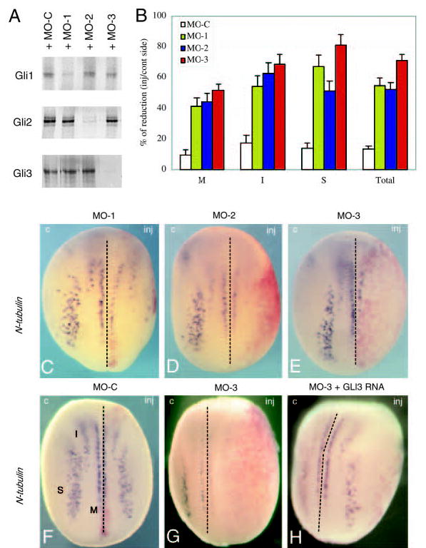
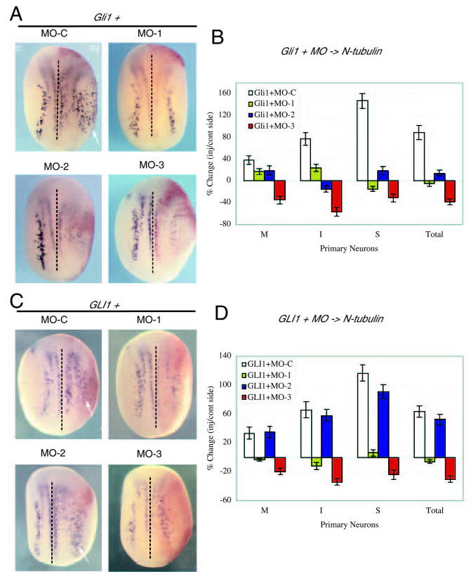
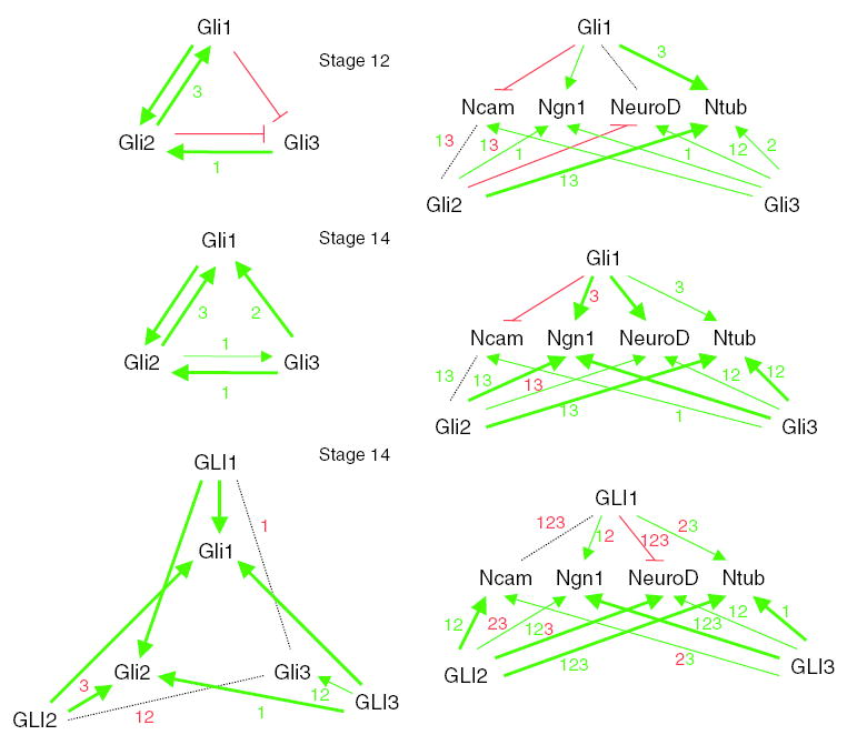
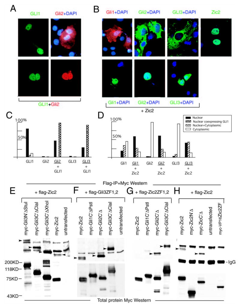
The Gli proteins are critical components of multiple processes in development, homeostasis and disease, including neurogenesis and tumorigenesis. However, it is unclear how the Gli code, the sum of their combinatorial positive and negative functions, dictates cell fate and behavior. Using an antisense approach to knockdown gene function in vivo, we find that each of the three Gli proteins is required for the induction of all primary neurons in the amphibian neural plate and regulates the bHLH/Notch neurogenic cascade. Analyses of endogenous Gli function in Gli-mediated neurogenesis and tumorigenesis, and in animal cap assays, reveal specific requirements that are context specific. Nuclear colocalization and binding studies suggest the formation of complexes, with the first two zinc fingers of the Gli five zinc-finger domain acting as a protein-protein interaction site. The Gli proteins therefore appear to form a dynamic physical network that underlies cooperative function, greatly extending the combinatorial possibilities of the Gli code, which may be further fine-tuned in cell fate specification by co-factor function.
Figures


Similar articles
-
Gli/Zic factors pattern the neural plate by defining domains of cell differentiation.Nature. 1998 Jun 11;393(6685):579-83. doi: 10.1038/31242. Nature. 1998. PMID: 9634234
-
Xmeis1, a protooncogene involved in specifying neural crest cell fate in Xenopus embryos.Oncogene. 2001 Mar 15;20(11):1329-42. doi: 10.1038/sj.onc.1204250. Oncogene. 2001. PMID: 11313877
-
Gli1 is a target of Sonic hedgehog that induces ventral neural tube development.Development. 1997 Jul;124(13):2537-52. doi: 10.1242/dev.124.13.2537. Development. 1997. PMID: 9216996
-
Gli genes and limb development.Cell Tissue Res. 1999 Apr;296(1):75-83. doi: 10.1007/s004410051268. Cell Tissue Res. 1999. PMID: 10199967 Review.
-
Catching a Gli-mpse of Hedgehog.Cell. 1997 Jul 25;90(2):193-6. doi: 10.1016/s0092-8674(00)80325-6. Cell. 1997. PMID: 9244291 Review. No abstract available.
Cited by
-
Members of the Rusc protein family interact with Sufu and inhibit vertebrate Hedgehog signaling.Development. 2016 Nov 1;143(21):3944-3955. doi: 10.1242/dev.138917. Epub 2016 Sep 15. Development. 2016. PMID: 27633991 Free PMC article.
-
Genome-wide microarray expression and genomic alterations by array-CGH analysis in neuroblastoma stem-like cells.PLoS One. 2014 Nov 13;9(11):e113105. doi: 10.1371/journal.pone.0113105. eCollection 2014. PLoS One. 2014. PMID: 25392930 Free PMC article.
-
cDNA microarray gene expression profiling of hedgehog signaling pathway inhibition in human colon cancer cells.PLoS One. 2010 Oct 1;5(10):e13054. doi: 10.1371/journal.pone.0013054. PLoS One. 2010. PMID: 20957031 Free PMC article.
-
Cerebellar development and disease.Curr Opin Neurobiol. 2008 Feb;18(1):12-9. doi: 10.1016/j.conb.2008.05.010. Epub 2008 May 29. Curr Opin Neurobiol. 2008. PMID: 18513948 Free PMC article. Review.
-
A Retinoic Acid-Hedgehog Cascade Coordinates Mesoderm-Inducing Signals and Endoderm Competence during Lung Specification.Cell Rep. 2016 Jun 28;16(1):66-78. doi: 10.1016/j.celrep.2016.05.060. Epub 2016 Jun 16. Cell Rep. 2016. PMID: 27320915 Free PMC article.
References
-
- Agren M, Kogerman P, Kleman MI, Wessling M, Toftgard R. Expression of the PTCH1 tumor suppressor gene is regulated by alternative promoters and a single functional Gli-binding site. Gene. 2004;330:101–114. - PubMed
-
- Aza-Blanc P, Ramirez-Weber FA, Laget MP, Schwartz C, Kornberg TB. Proteolysis that is inhibited by hedgehog targets Cubitus interruptus protein to the nucleus and converts it to a represser. Cell. 1997;89:1043–1053. - PubMed
-
- Aza-Blanc P, Lin HY, Ruiz i Altaba A, Kornberg TB. Expression of the vertebrate Gli proteins in Drosophila reveals a distribution of activator and represser activities. Development. 2000;127:4293–4301. - PubMed
-
- Bai CB, Joyner AL. Gli1 can rescue the in vivo function of Gli2. Development. 2001;128:5161–5172. - PubMed
-
- Bai CB, Auerbach W, Lee JS, Stephen D, Joyner AL. Gli2, but not Gli1, is required for initial Shh signaling and ectopic activation of the Shh pathway. Development. 2002;129:475347–475361. - PubMed
Publication types
MeSH terms
Substances
Grants and funding
LinkOut - more resources
Full Text Sources
Other Literature Sources
Miscellaneous
