. 2005;4(2):9.
doi: 10.1186/jbiol24. Epub 2005 Jun 27.

Guanine-nucleotide exchange on ribosome-bound elongation factor G initiates the translocation of tRNAs

Affiliations

Guanine-nucleotide exchange on ribosome-bound elongation factor G initiates the translocation of tRNAs

Andrey V Zavialov et al. J Biol. 2005.

Abstract

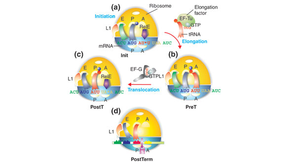
Background: During the translation of mRNA into polypeptide, elongation factor G (EF-G) catalyzes the translocation of peptidyl-tRNA from the A site to the P site of the ribosome. According to the 'classical' model, EF-G in the GTP-bound form promotes translocation, while hydrolysis of the bound GTP promotes dissociation of the factor from the post-translocation ribosome. According to a more recent model, EF-G operates like a 'motor protein' and drives translocation of the peptidyl-tRNA after GTP hydrolysis. In both the classical and motor protein models, GDP-to-GTP exchange is assumed to occur spontaneously on 'free' EF-G even in the absence of a guanine-nucleotide exchange factor (GEF).

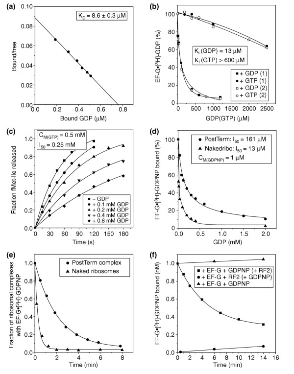
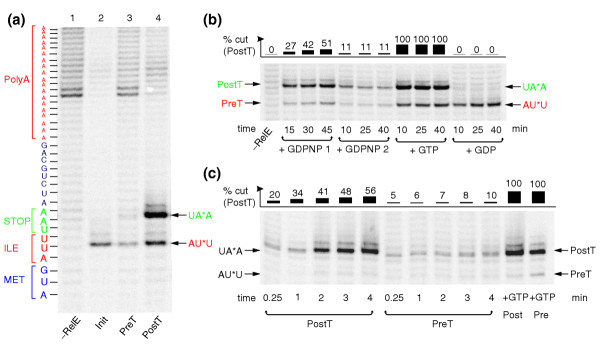
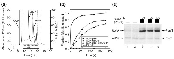
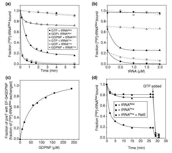
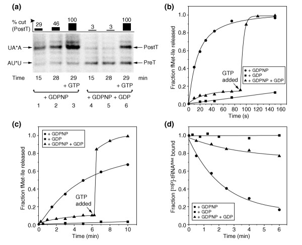
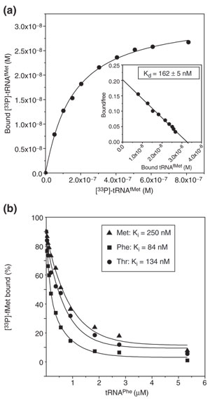
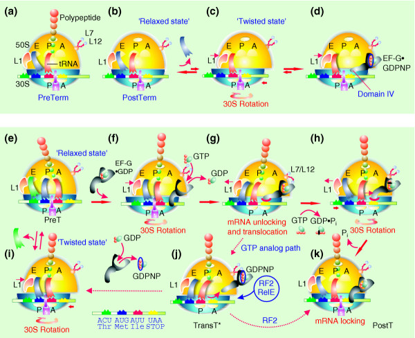
Results: We have made a number of findings that challenge both models. First, free EF-G in the cell is likely to be in the GDP-bound form. Second, the ribosome acts as the GEF for EF-G. Third, after guanine-nucleotide exchange, EF-G in the GTP-bound form moves the tRNA2-mRNA complex to an intermediate translocation state in which the mRNA is partially translocated. Fourth, subsequent accommodation of the tRNA2-mRNA complex in the post-translocation state requires GTP hydrolysis.

Conclusion: These results, in conjunction with previously published cryo-electron microscopy reconstructions of the ribosome in various functional states, suggest a novel mechanism for translocation of tRNAs on the ribosome by EF-G. Our observations suggest that the ribosome is a universal guanosine-nucleotide exchange factor for EF-G as previously shown for the class-II peptide-release factor 3.


Figures

Figure 1
Figure 1
Schematic representation of (a) initiation, (b) pre-translocation, (c) post-translocation, and (d) post-termination complexes, referred to as Init, preT and postT, and postTerm, respectively. A, amino-acyl tRNA site on the ribosome; P, peptidyl-tRNA site; E, exit site; L1, ribosomal protein. The large subunit of the ribosome is shown in yellow and the small subunit in blue. The colored ribbons represent tRNAs and the colored balls represent amino acids in aminoacyl- or peptidyl-tRNA. The purple arrow represents RelE, which cleaves the codon shown at the *. The mauve padlock in (d) illustrates a state of the ribosome in which the mRNA is locked, and cannot move in relation to the small subunit. The figure represents a special case in which the postT ribosome has a stop codon (UAA) in the A site, and is therefore also a pre-termination (preTerm) ribosome. For further details see text and Figure 8.
Figure 2
Figure 2
Ribosome-dependent exchange of GDP to GTP on EF-G. (a) Scatchard plot from a nitrocellulose-filtration experiment to obtain the dissociation constant for the binding of [3H]-GDP to free EF-G. (b) Chase of [3H]-GDP from free EF-G by unlabeled GTP or, as a control, GDP. The dissociation constant for GTP binding to free EF-G was obtained from the corresponding constant for GDP binding in (a) and from the inhibition of [3H]-GDP binding to EF-G by GTP addition. The figure shows the results of two independent experiments (1 and 2). (c) Time-dependent release of fMet-Ile by 0.5 μM RF2 after translocation of fMet-Ile-tRNAIle from the A to the P site by a catalytic amount of EF-G (10 nM) added to 23 nM preT ribosomes together with 0.5 mM GTP and 0–0.8 mM GDP. CM(GTP) is the GTP concentration and I50 is the GDP concentration at which the rate of translocation is reduced to half-maximal value. (d) Inhibition of EF-G•GDPNP binding to post-termination (PostTerm) complexes or naked 70S ribosomes (Nakedribo) in the presence of 1 μM [3H]-GDPNP and 0–2 mM unlabeled GDP. (e) Fraction of [3H]-GDPNP (total concentration 1 μM) bound to EF-G• [3H]-GDPNP in postTerm complexes or in naked ribosomes as a function of time after addition of unlabeled GDP to a concentration of 2 mM. (f) Time-dependence of EF-G• [3H]-GDPNP binding to postTerm ribosomes in the presence of 1 μM [3H]-GDPNP: in the absence of RF2 (control), after addition of [3H]-GDPNP to EF-G pre-incubated with RF2 and postTerm ribosomes, or after addition of RF2 to EF-G pre-incubated with [3H]-GDPNP and postTerm ribosomes.
Figure 3
Figure 3
RelE cleavage of mRNA in the A site of ribosomal complexes. (a) The mRNA fragments resulting from RelE cleavage in the A site of the three ribosomal complexes Init (see Figure 1a), preT (see Figure 1b) and postT (see Figure 1c), separated on a 10% sequencing gel. The amount of radioactivity in the postT lane was doubled to make the AUU cleavage visible. (b) Time-dependent cleavage of mRNA by RelE; preT ribosomes were incubated with EF-G together with GDPNP (+ GDPNP 2) or GTP (+ GTP) or GDP (+ GDP). RelE was added after 10, 25 or 40 min, and the reaction was in each case quenched 5 min after RelE addition. Alternatively, preT ribosomes were incubated together with EF-G, RelE and GDPNP and the reaction quenched after 15, 30 or 45 min (+ GDPNP 1). (c) Time-dependent cleavage of mRNA by 120 nM RelE in the A site of 0.3 μM postT or preT ribosome complexes incubated with 2 μM EF-G and 0.6 mM GDPNP. As a control, in the last two lanes 1 mM GTP was added to postT or preT ribosomes at the end of the incubation.
Figure 4
Figure 4
Contamination of GDP preparations with GTP strongly stimulates translocation by EF-G. (a) Elution profile of commercially available GDP from a MonoQ column showing the GTP and GMP contaminations. %B is the percentage of buffer B (20 mM Tris-HCl, 1 M NaCl) in the buffer A (20 mM Tris-HCl) + B mixture. (b) Time-dependent release of peptide by 0.4 μM RF2 after translocation of fMet-Ile-tRNA (23 nM total) from the A site to the P site by 1 μM EF-G in the presence of 1 mM purified GDP, unpurified GDP, purified GDP containing 20 μM GTP (2%), or 20 μM GTP. (c) Cleavage of mRNA by RelE incubated with 0.15 μM preT, 2 μM EF-G and nucleotides. Lanes: (1) no GDP; (2) 1 mM purified GDP; (3) 1 mM unpurified GDP; (4) 1 mM purified GDP containing 2% GTP; (5) 20 μM GTP.
Figure 5
Figure 5
Properties of the transition state. (a) Time-dependent exchange of [33P]-tRNAfMet bound to the P site of 70 nM preT complex with 1 μM unlabeled tRNAfMet or tRNAPhe after the addition of 2 μM EF-G and 1 mM nucleotide. (b) The fraction of [33P]-tRNAfMet exchanged with tRNAfMet or tRNAPhe after 9 min incubation of 70 nM preT with 2 μM EF-G, 1 mM nucleotide and 0–2 μM tRNAfMet or tRNAPhe. (c) Fraction of [33P]-tRNAfMet on 88 nM preT ribosomes exchanged after 7 min incubation with 2 μM unlabeled tRNAfMet, 2 μM EF-G and 0–240 μM GDPNP to estimate the fraction of ribosomes containing EF-G•GDPNP. (d) Exchange of [33P]-tRNAfMet with 2 μM tRNAfMet or tRNAPhe added to 78 nM preT incubated with 2 μM EF-G, 0.4 nM GDPNP with or without 80 nM RelE. At 27.5 min, 1 mM GTP was added to translocate [33P]-tRNA fMet to the E site.
Figure 6
Figure 6
Removal of EF-G•GDPNP from the transition state with GDP. (a) Time-dependent cleavage of mRNA by 166 nM RelE in transT* complex in the presence of 2 μM EF-G and 0.32 mM GDPNP (GDPNP case) or after further addition of GDP to a concentration of 1 mM to remove EF-G from the ribosome (GDPNP + GDP case). In each case, GTP was added to a final concentration of 1 mM at 29 min to show the fraction of ribosomes that was active in translocation (lanes 3 and 6). (b,c) Time-dependent release of fMet-Ile by (b) 0.4 mM puromycin or (c) 0.5 μM RF2; 2 μM EF-G was pre-incubated with 46 nM preT complex and 40 μM GDPNP or polymix buffer for 3 min at 37°C. Then, buffer or 2 mM GDP was added and the incubation was continued for 1 min. Finally, (b) 0.4 mM puromycin or (c) 0.5 μM RF2 was added and the extent of peptide release was observed over time. (d) Exchange of [33P]-tRNAfMet on 88 nM preT complex, pre-incubated with 2 μM EF-G and 100 μM GDPNP or with buffer, with 2 μM tRNAfMet in the presence or absence of 2 mM GDP.
Figure 7
Figure 7
Binding of deacylated tRNA to the E site. (a) Binding of [33P]-tRNAfMet to the postT complex with fMet-Phe-Ile-tRNAIlein the P site and a Thr codon (ACG) in the E site. Insert: Scatchard plot to obtain the dissociation constant. (b) Chase of [33P]-tRNAfMet from the E site of postT complexes containing Met (AUG), Phe (UUU) or Thr (ACG) codons with unlabeled tRNAPhe. Dissociation constants for [33P] tRNAfMet were obtained as in (a) and the dissociation constants for tRNAPhe were calculated from the 50% chase (I50) concentrations of tRNAPhe.
Figure 8
Figure 8
Interaction between the ribosome and EF-G; a proposal for the whole mechanism of translocation. (a-c) Release of peptide from the postTerm ribosome allows the intersubunit rotation. (d) EF-G•GDPNP stabilizes the twisted form of the ribosome as observed by cryo-EM [5]. (e-h,k) A model explaining the mechanism of translocation. (i,j) A GTP-analog pathway. The model is explained in detail in the text and symbols are as in Figure 1. L7/12 is a complex of ribosomal proteins thought to activate GTP hydrolysis on ribosomal GTPases. The mauve padlock illustrates states of the ribosome in which the mRNA is locked, and cannot move in relation to the small subunit. Domain IV of EF-G is suggested in the main text to play an important role in translocation.

References

    1. Bourne HR, Sanders DA, McCormick F. The GTPase superfamily: conserved structure and molecular mechanism. Nature. 1991;349:117–127. doi: 10.1038/349117a0. - DOI - PubMed
    1. Sprang SR. G protein mechanisms: insights from structural analysis. Annu Rev Biochem. 1997;66:639–678. doi: 10.1146/annurev.biochem.66.1.639. - DOI - PubMed
    1. Savelsbergh A, Mohr D, Wilden B, Wintermeyer W, Rodnina MV. Stimulation of the GTPase activity of translation elongation factor G by ribosomal protein L7/12. J Biol Chem. 2000;275:890–894. doi: 10.1074/jbc.275.2.890. - DOI - PubMed
    1. Czworkowski J, Moore PB. The conformational properties of elongation factor G and the mechanism of translocation. Biochemistry. 1997;36:10327–10334. doi: 10.1021/bi970610k. - DOI - PubMed
    1. Valle M, Zavialov A, Sengupta J, Rawat U, Ehrenberg M, Frank J. Locking and unlocking of ribosomal motions. Cell. 2003;114:123–134. doi: 10.1016/S0092-8674(03)00476-8. - DOI - PubMed

Publication types

MeSH terms

Substances

LinkOut - more resources