. 2005 Jun 28:5:9.
doi: 10.1186/1472-6807-5-9.

An inactivated nuclease-like domain in RecC with novel function: implications for evolution

Affiliations

An inactivated nuclease-like domain in RecC with novel function: implications for evolution

Daniel John Rigden. BMC Struct Biol. .

Abstract

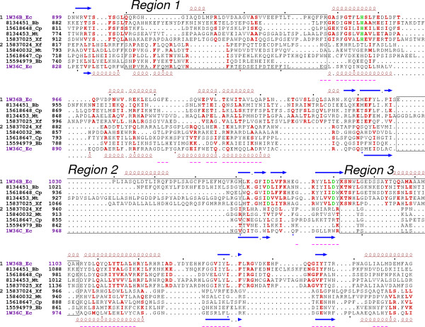
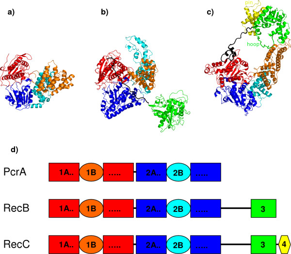
Background: The PD-(D/E)xK superfamily, containing a wide variety of other exo- and endonucleases, is a notable example of general function conservation in the face of extreme sequence and structural variation. Almost all members employ a small number of shared conserved residues to bind catalytically essential metal ions and thereby effect DNA cleavage. The crystal structure of the RecBCD prokaryotic DNA repair machinery shows that RecB contains such a nuclease domain at its C-terminus. The RecC C-terminal region was reported as having a novel fold.

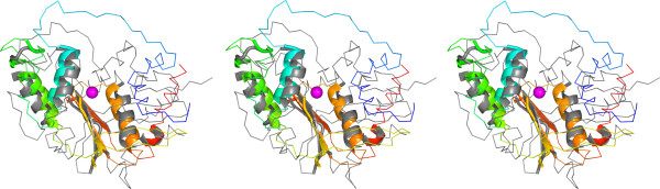
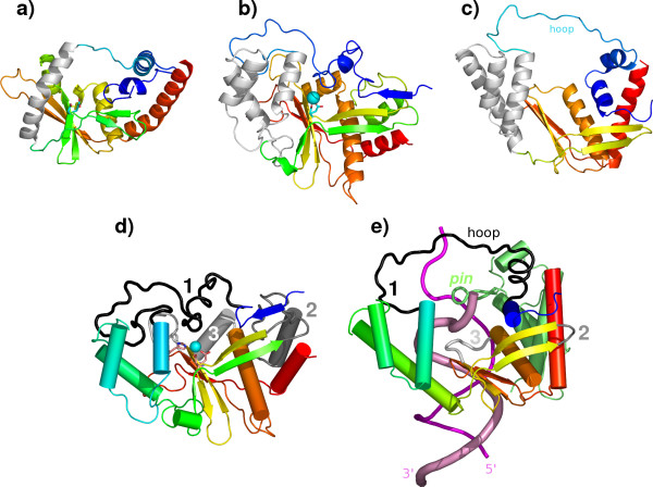
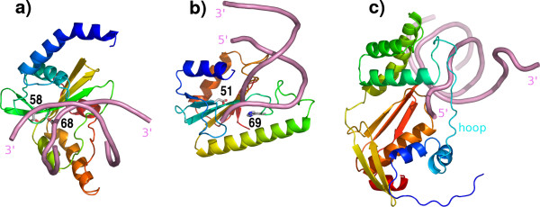
Results: The RecC C-terminal region can be divided into an alpha/beta domain and a smaller alpha-helical bundle domain. Here we show that the alpha/beta domain is homologous to the RecB nuclease domain but lacks the features necessary for catalysis. Instead, the domain has a novel function within the nuclease superfamily--providing a hoop through which single-stranded DNA passes. Comparison with other structures of nuclease domains bound to DNA reveals strikingly different modes of ligand binding. The alpha-helical bundle domain contributes the pin which splits the DNA duplex.

Conclusion: The demonstrated homology of RecB and RecC shows how evolution acted to produce the present RecBCD complex through aggregation of new domains as well as functional divergence and structural redeployment of existing domains. Distantly homologous nuclease(-like) domains bind DNA in highly diverse manners.


Figures

Figure 1
Figure 1
Stereo structural superposition of the nuclease(-like) domains of RecB and RecC produced with LSQMAN [29]. RecB is coloured grey while RecC, is coloured in a spectrum from blue (N-terminus) to red (C-terminus). Structurally superposed regions are shown in cartoon representation (rms deviation of 2.19Å for 71 Cα atoms), other parts as a Cα trace. The calcium ion bound to RecB is shown as a magenta sphere.
Figure 2
Figure 2
Comparisons of structurally aligned nuclease(-like) domains in λ-exonuclease, RecB and RecC. The comparison in a)-c) shows how a single helix in λ-exonuclease (PDB code 1avq; [5]) (a) has been replaced by superimposable α-helical bundles in RecB (b) and RecC (c) (PDB code 1w36; [18]), indicating a more recent shared ancestor of the latter pair. The regions in question are shown as light grey. The remainders of the molecules are coloured in a spectrum from blue (N-terminus) to red (C-terminus). In a) and b), two acidic, metal-ligating residues drawn as sticks mark respective catalytic sites. RecB and RecC are compared in more detail in d) and e), respectively, again coloured from blue to red with the exception of labelled key regions 1 (black), 2 (dark grey) and 3 (grey). Bound metal is shown in b) and d) as spheres while e) additionally shows DNA (shades of pink) and the domain 4 of RecC, coloured uniformly lime green with its pin structure labelled. The DNA strand that penetrates the hoop provided by RecC is shown as a broader cartoon. The RecC "hoop" region (see text for details) is labelled in c) and e) and DNA strand termini are labelled in e).
Figure 3
Figure 3
Structure-based sequence alignment of the nuclease(-like) domains of RecB and RecC. Nuclease(-like) domain sequences of RecB (above, group 1) and RecC (below, group 2) were chosen from diverse representative species and extracted from complete alignments of COG database [31] entries for RecB or RecC. Purple indicates the E. coli sequences crystallized as the RecBCD complex (PDB code 1w36; [18]). Other sequences are labelled with Genbank numbers and sequence codes Bb, Borrelia burgdorferi; Cp, Chlamydophila pneumoniae; Mt, Mycobacterium tuberculosis; Xf, Xylella fastidiosa. Red colouring indicates conservation within each group while green is used for three important catalytic residues of RecB – H956, D1067 and D1080 [18]. Elements of regular secondary structure are shown above (RecB) and below (RecC) the alignment, where spirals represent α-helices and arrows β-strands. The three key regions (numbered 1–3) involved in adaptation of the RecC nuclease-like domain to its new function, as discussed in the text (see also Fig 2), are boxed and labelled. Purple underlining indicates zones that can be simultaneously structurally aligned (rms deviation of 2.19Å for 71 Cα atoms).
Figure 4
Figure 4
Comparison of modes of DNA binding to superimposed nuclease(-like) domains. The domain structures are those of a) PvuII (PDB code 1pvi; [22] b) vsr exonuclease (PDB code 1odg; [23]) and c) RecC (PDB code 1w36; [18]). Protein chains are coloured in a spectrum from blue (N-terminus) to red (C-terminus) while DNA is coloured uniformly pink. In order to illustrate the approximate locations of the catalytic sites, selected catalytic residues are shown for PvuII (D58 and E58) and vsr exonuclease (D51 and H69). DNA termini are labelled, as is the "hoop" in RecC.
Figure 5
Figure 5
Domain comparison of PcrA, RecB and RecC. The structures of a) substrate-complexed PcrA (PDB code 3pjr; [38]), b) RecB and c) RecC (PDB code 1w36; [18]), are superimposed using domain 2A. The domains 1A are coloured red, while orange is used for 1B, blue for 2A, cyan for 2B, green for 3 and yellow for 4. The same colours are used in the schematic diagram d) which illustrates how evolution progressed through addition of domains. The "pin" and "hoop" in RecC (see text for details) are labelled. In PcrA the residues defining the starts of the various (sub-)domains are 1B, 92; 1A continuation, 218; 2A, 287; 2B, 385; 2A continuation, 553. In RecB the corresponding residue numbers are 1B, 151; 1A continuation, 349; 2A, 446; 2B, 583; 2A continuation, 729; linker, 870; 3, 900. In RecC they are 1B, 79; 1A continuation, 208; 2A, 329; 2B, 443; 2A continuation, 649; linker, 784; 3, 828; 4, 1034.

Similar articles

Cited by

References

    1. Aggarwal AK. Structure and function of restriction endonucleases. Curr Opin Struct Biol. 1995;5:11–19. doi: 10.1016/0959-440X(95)80004-K. - DOI - PubMed
    1. Tsutakawa SE, Muto T, Kawate T, Jingami H, Kunishima N, Ariyoshi M, Kohda D, Nakagawa M, Morikawa K. Crystallographic and functional studies of very short patch repair endonuclease. Mol Cell. 1999;3:621–628. doi: 10.1016/S1097-2765(00)80355-X. - DOI - PubMed
    1. Hickman AB, Li Y, Mathew SV, May EW, Craig NL, Dyda F. Unexpected structural diversity in DNA recombination: the restriction endonuclease connection. Mol Cell. 2000;5:1025–1034. doi: 10.1016/S1097-2765(00)80267-1. - DOI - PubMed
    1. Bond CS, Kvaratskhelia M, Richard D, White MF, Hunter WN. Structure of Hjc, a Holliday junction resolvase, from Sulfolobus solfataricus. Proc Natl Acad Sci U S A. 2001;98:5509–5514. doi: 10.1073/pnas.091613398. - DOI - PMC - PubMed
    1. Kovall R, Matthews BW. Toroidal structure of lambda-exonuclease. Science. 1997;277:1824–1827. doi: 10.1126/science.277.5333.1824. - DOI - PubMed

MeSH terms

LinkOut - more resources